


Vol 7, No 2 (2015)
- Year: 2015
- Published: 15.06.2015
- Articles: 14
- URL: https://actanaturae.ru/2075-8251/issue/view/837

Reviews
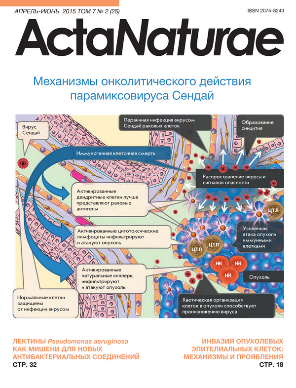
Mechanisms of Oncolysis by Paramyxovirus Sendai
Abstract
Some viral strains of the Paramyxoviridae family may be used as anti-tumor agents. Oncolytic paramyxoviruses include attenuated strains of the measles virus, Newcastle disease virus, and Sendai virus. These viral strains, and the Sendai virus in particular, can preferentially induce the death of malignant, rather than normal, cells. The death of cancer cells results from both direct killing by the virus and through virus-induced activation of anticancer immunity. Sialic-acid-containing glycoproteins that are overexpressed in cancer cells serve as receptors for some oncolytic paramyxoviruses and ensure preferential interaction of paramyxoviruses with malignant cells. Frequent genetic defects in interferon and apoptotic response systems that are common to cancer cells ensure better susceptibility of malignant cells to viruses. The Sendai virus as a Paramyxovirus is capable of inducing the formation of syncytia, multinuclear cell structures which promote viral infection spread within a tumor without virus exposure to host neutralizing antibodies. As a result, the Sendai virus can cause mass killing of malignant cells and tumor destruction. Oncolytic paramyxoviruses can also promote the immune-mediated elimination of malignant cells. In particular, they are powerful inducers of interferon and other cytokynes promoting antitumor activity of various cell components of the immune response, such as dendritic and natural killer cells, as well as cytotoxic T lymphocytes. Taken together these mechanisms explain the impressive oncolytic activity of paramyxoviruses that hold promise as future, efficient anticancer therapeutics.
 6-16
6-16


Cancer Invasion: Patterns and Mechanisms
Abstract
Cancer invasion and the ability of malignant tumor cells for directed migration and metastasis have remained a focus of research for many years. Numerous studies have confirmed the existence of two main patterns of cancer cell invasion: collective cell migration and individual cell migration, by which tumor cells overcome barriers of the extracellular matrix and spread into surrounding tissues. Each pattern of cell migration displays specific morphological features and the biochemical/molecular genetic mechanisms underlying cell migration. Two types of migrating tumor cells, mesenchymal (fibroblast-like) and amoeboid, are observed in each pattern of cancer cell invasion. This review describes the key differences between the variants of cancer cell migration, the role of epithelial-mesenchymal, collective-amoeboid, mesenchymal-amoeboid, and amoeboid-mesenchymal transitions, as well as the significance of different tumor factors and stromal molecules in tumor invasion. The data and facts collected are essential to the understanding of how the patterns of cancer cell invasion are related to cancer progression and therapy efficacy. Convincing evidence is provided that morphological manifestations of the invasion patterns are characterized by a variety of tissue (tumor) structures. The results of our own studies are presented to show the association of breast cancer progression with intratumoral morphological heterogeneity, which most likely reflects the types of cancer cell migration and results from different activities of cell adhesion molecules in tumor cells of distinct morphological structures.
17-28
Pseudomonas Aeruginosa Lectins As Targets for Novel Antibacterials
Abstract
Pseudomonas aeruginosa is one of the most widespread and troublesome opportunistic pathogens that is capable of colonizing various human tissues and organs and is often resistant to many currently used antibiotics. This resistance is caused by different factors, including the acquisition of specific resistance genes, intrinsic capability to diminish antibiotic penetration into the bacterial cell, and the ability to form biofilms. This situation has prompted the development of novel compounds differing in their mechanism of action from traditional antibiotics that suppress the growth of microorganisms or directly kill bacteria. Instead, these new compounds should decrease the pathogens’ ability to colonize and damage human tissues by inhibiting the virulence factors and biofilm formation. The lectins LecA and LecB that bind galactose and fucose, as well as oligo- and polysaccharides containing these sugars, are among the most thoroughly-studied targets for such novel antibacterials. In this review, we summarize the results of experiments highlighting the importance of these proteins for P. aeruginosa pathogenicity and provide information on existing lectins inhibitors and their effectiveness in various experimental models. Particular attention is paid to the effects of lectins inhibition in animal models of infection and in clinical practice. We argue that lectins inhibition is a perspective approach to combating P. aeruginosa. However, despite the existence of highly effective in vitro inhibitors, further experiments are required in order to advance these inhibitors into pre-clinical studies.
29-41
NeuN As a Neuronal Nuclear Antigen and Neuron Differentiation Marker
Abstract
The NeuN protein is localized in nuclei and perinuclear cytoplasm of most of the neurons in the central nervous system of mammals. Monoclonal antibodies to the NeuN protein have been actively used in the immunohistochemical research of neuronal differentiation to assess the functional state of neurons in norm and pathology for more than 20 years. Recently, NeuN antibodies have begun to be applied in the differential morphological diagnosis of cancer. However, the structure of the protein, which can be revealed by antibodies to NeuN, remained unknown until recently, and the functions of the protein are still not fully clear. In the present mini-review, data on NeuN accumulated so far are summarized and analyzed. Data on the structure and properties of the protein, its isoforms, intracellular localization, and hypothesized functions are reported. The application field of immunocytochemical detection of NeuN in scientific and clinical studies, as well as the difficulties in the interpretation of the obtained experimental data and their possible causes, is described in details.
42-47
In Vitro Mouse Ovarian Follicle Growth and Maturation in Alginate Hydrogel: Current State of the Art
Abstract
This review describes the main factors affecting the in vitro development of mouse ovarian follicles under conditions of three-dimensional alginate hydrogel system. The factors discussed include concentration of alginate hydrogel, presence of additives (collagen, fibrin) influencing substrate rigidity; culture conditions; composition of culture media; substances that act like antioxidants (salts of ascorbic acid, glutathione) and contribute to the improvement of lipid metabolism (L-carnitine), hormones and growth factors. The methods for follicle group cultivation in alginate hydrogel and cocultivation of different cell populations with follicles encapsulated in alginate hydrogel are covered in the present article.
48-56
Research Articles
Search for Human Lactate Dehydrogenase A Inhibitors Using Structure-Based Modeling
Abstract
The human lactate dehydrogenase isoform A plays an important role in the anaerobic metabolism of tumour cells and therefore constitutes an attractive target in the oncology field. Full-atom models of lactate dehydrogenase A (in complex with NADH and in the apo form) have been generated to enable structure-based design of novel inhibitors competing with pyruvate and NADH. The structural criteria for the selection of potential inhibitors were established, and virtual screening of a library of low-molecular-weight compounds was performed. A potential inhibitor, STK381370, was identified whose docking pose was stabilized through additional interactions with the loop 96-111 providing for the transition from the open to the closed conformation.
57-63
Modified Method of rRNA Structure Analysis Reveals Novel Characteristics of Box C/D RNA Analogues
Abstract
Ribosomal RNA (rRNA) maturation is a complex process that involves chemical modifications of the bases or sugar residues of specific nucleotides. One of the most abundant types of rRNA modifications, ribose 2’-O-methylation, is guided by ribonucleoprotein complexes containing small nucleolar box C/D RNAs. Since the majority of 2’-O-methylated nucleotides are located in the most conserved regions of rRNA that comprise functionally important centers of the ribosome, an alteration in a 2’-O-methylation profile can affect ribosome assembly and function. One of the key approaches for localization of 2’-O-methylated nucleotides in long RNAs is a method based on the termination of reverse transcription. The current study presents an adaptation of this method for the use of fluorescently labeled primers and analysis of termination products by capillary gel electrophoresis on an automated genetic analyzer. The developed approach allowed us to analyze the influence of the synthetic analogues of box C/D RNAs on post-transcriptional modifications of human 28S rRNA in MCF-7 cells. It has been established that the transfection of MCF-7 cells with a box C/D RNA analogue leads to an enhanced modification level of certain native sites of 2’-O-methylation in the target rRNA. The observed effect of synthetic RNAs on the 2’-O-methylation of rRNA in human cells demonstrates a path towards targeted regulation of rRNA post-transcriptional maturation. The described approach can be applied in the development of novel diagnostic methods for detecting diseases in humans.
64-73
Specific Depletion of Myelin-Reactive B Cells via BCR-Targeting
Abstract
B cells play a crucial role in the development and pathogenesis of systemic and organ-specific autoimmune diseases. Autoreactive B cells not only produce antibodies, but also secrete pro-inflammatory cytokines and present specific autoantigens to T cells. The treatment of autoimmune diseases via the elimination of the majority of B cells using the monoclonal anti-CD19/20 antibody (Rituximab) causes systemic side effects and, thus, requires a major revision. Therapeutic intervention directed towards selective elimination of pathogenic autoreactive B cells has the potential to become a universal approach to the treatment of various autoimmune abnormalities. Here, we developed a recombinant immunotoxin based on the immunodominant peptide of the myelin basic protein (MBP), fused to the antibody Fc domain. We showed that the obtained immunotoxin provides selective in vivo elimination of autoreactive B cells in mice with experimental autoimmune encephalomyelitis. The proposed conception may be further used for the development of new therapeutics for a targeted treatment of multiple sclerosis and other autoimmune disorders.
74-79
Determination of Alkali-Sensing Parts of the Insulin Receptor-Related Receptor Using the Bioinformatic Approach
Abstract
IRR (insulin receptor-related receptor) is a receptor tyrosine kinase belonging to the insulin receptor family, which also includes insulin receptor and IGF-IR receptor. We have previously shown that IRR is activated by extracellular fluid with pH > 7.9 and regulates excess alkali excretion in the body. We performed a bioinformatic analysis of the pH-sensitive potential of all three members of the insulin receptor family of various animal species (from frog to man) and their chimeras with swapping of different domains in the extracellular region. An analysis using the AcalPred program showed that insulin receptor family proteins are divided into two classes: one class with the optimal working pH in the acidic medium (virtually all insulin receptor and insulin-like growth factor receptor orthologs, except for the IGF-IR ortholog from Xenopus laevis) and the second class with the optimal working pH in the alkaline medium (all IRR orthologs). The program had predicted that the most noticeable effect on the pH-sensitive property of IRR would be caused by the replacement of the L1 and C domains in its extracellular region, as well as the replacement of the second and third fibronectin repeats. It had also been assumed that replacement of the L2 domain would have the least significant effect on the alkaline sensitivity of IRR. To test the in silico predictions, we obtained three constructs with swapping of the L1C domains, the third L2 domain, and all three domains L1CL2 of IRR with similar domains of the insulin-like growth factor receptor. We found that replacement of the L1C and L1CL2 domains reduces the receptor’s ability to be activated with alkaline pH, thus increasing the half-maximal effective concentration by about 100%. Replacement of the L2 domain increased the half-maximal effective concentration by 40%. Thus, our results indicate the high predictive potential of the AcalPred algorithm, not only for the pH-sensitive enzymes, but also for pH-sensitive receptors.
80-86
Detection of T-Cadherin Expression in Mouse Embryos
Abstract
The aim of the present study was to evaluate T-cadherin expression at the early developmental stages of the mouse embryo. Using in situ hybridization and immunofluorescent staining of whole embryos in combination with confocal microscopy, we found that T-cadherin expression is detected in the developing brain, starting with the E8.75 stage, and in the heart, starting with the E11.5 stage. These data suggest a possible involvement of T-cadherin in the formation of blood vessels during embryogenesis.
87-94
The Effect of Hydrophobic Monoamines on Acid-Sensing Ion Channels ASIC1B
Abstract
Acid-sensing ion channels (ASICs) are widely distributed in both the central and peripheral nervous systems of vertebrates. The pharmacology of these receptors remains poorly investigated, while the search for new ASIC modulators is very important. Recently, we found that some monoamines, which are blockers of NMDA receptors, inhibit and/or potentiate acid-sensing ion channels, depending on the subunit composition of the channels. The effect of 9-aminoacridine, IEM-1921, IEM-2117, and memantine both on native receptors and on recombinant ASIC1a, ASIC2a, and ASIC3 homomers was studied. In the present study, we have investigated the effect of these four compounds on homomeric ASIC1b channels. Experiments were performed on recombinant receptors expressed in CHO cells using the whole-cell patch clamp technique. Only two compounds, 9-aminoacridine and memantine, inhibited ASIC1b channels. IEM-1921 and IEM-2117 were inactive even at a 1000 μM concentration. In most aspects, the effect of the compounds on ASIC1b was similar to their effect on ASIC1a. The distinguishing feature of homomeric ASIC1b channels is a steep activation-dependence, indicating cooperative activation by protons. In our experiments, the curve of the concentration dependence of ASIC1b inhibition by 9-aminoacridine also had a slope (Hill coefficient) of 3.8, unlike ASIC1a homomers, for which the Hill coefficient was close to 1. This finding indicates that the inhibitory effect of 9-aminoacridine is associated with changes in the activation properties of acid-sensing ion channels.
95-101
Thio Derivatives of 2(5H)-Furanone As Inhibitors against Bacillus subtilis Biofilms
Abstract
Gram-positive bacteria cause a wide spectrum of infectious diseases, including nosocomial infections. While in the biofilm, bacteria exhibit increased resistance to antibiotics and the human immune system, causing difficulties in treatment. Thus, the development of biofilm formation inhibitors is a great challenge in pharmacology. The gram-positive bacterium Bacillus subtilis is widely used as a model organism for studying biofilm formation. Here, we report on the effect of new synthesized 2(5Н)-furanones on the biofilm formation by B.subtilis cells. Among 57 compounds tested, sulfur-containing derivatives of 2(5H)-furanone (F12, F15, and F94) repressed biofilm formation at a concentration of 10 μg/ml. Derivatives F12 and F94 were found to inhibit the biosynthesis of GFP from the promoter of the eps operon encoding genes of the biofilm exopolysaccharide synthesis (EPS). Using the differential fluorescence staining of alive/dead cells, we demonstrated an increased bacterial sensitivity to antibiotics (kanamycin and chloramphenicol) in the presence of F12, F15, and F94, with F12 being the most efficient one. The derivative F15 was capable of disrupting an already formed biofilm and thereby increasing the efficiency of antibiotics.
102-107
The Use of Atomic Force Microscopy for 3D Analysis of Nucleic Acid Hybridization on Microarrays
Abstract
Oligonucleotide microarrays are considered today to be one of the most efficient methods of gene diagnostics. The capability of atomic force microscopy (AFM) to characterize the three-dimensional morphology of single molecules on a surface allows one to use it as an effective tool for the 3D analysis of a microarray for the detection of nucleic acids. The high resolution of AFM offers ways to decrease the detection threshold of target DNA and increase the signal-to-noise ratio. In this work, we suggest an approach to the evaluation of the results of hybridization of gold nanoparticle-labeled nucleic acids on silicon microarrays based on an AFM analysis of the surface both in air and in liquid which takes into account of their three-dimensional structure. We suggest a quantitative measure of the hybridization results which is based on the fraction of the surface area occupied by the nanoparticles.
108-114
Redistribution of Free- and Cell-Surface-Bound DNA in Blood of Benign and Malignant Prostate Tumor Patients
Abstract
A direct correlation between the concentration of cell-free and cell-surface-bound circulating DNA (cfDNA and csbDNA, respectively) was demonstrated. Based on an inverse correlation between blood plasma DNase activity and the cfDNA concentration, blood DNases are supposed to regulate the cfDNA concentration. However, no correlation was found between the DNase activity in blood plasma and the csbDNA concentration, indicating that blood DNases are not involved in csbDNA dissociation from the cell surface. The possibility of DNA redistribution between cfDNA and csbDNA indicates that the total pool of circulating DNA (cfDNA +csbDNA) should be used for a correct analysis of marker DNA concentrations and data standardization.
115-118