Vol 13, No 3 (2021)
- Year: 2021
- Published: 15.11.2021
- Articles: 14
- URL: https://actanaturae.ru/2075-8251/issue/view/872
Reviews
Cardiovascular Effects of Snake Toxins: Cardiotoxicity and Cardioprotection
Abstract
Snake venoms, as complex mixtures of peptides and proteins, affect various vital systems of the organism. One of the main targets of the toxic components from snake venoms is the cardiovascular system. Venom proteins and peptides can act in different ways, exhibiting either cardiotoxic or cardioprotective effects. The principal classes of these compounds are cobra cardiotoxins, phospholipases A2, and natriuretic, as well as bradykinin-potentiating peptides. There is another group of proteins capable of enhancing angiogenesis, which include, e.g., vascular endothelial growth factors possessing hypotensive and cardioprotective activities. Venom proteins and peptides exhibiting cardiotropic and vasoactive effects are promising candidates for the design of new drugs capable of preventing or constricting the development of pathological processes in cardiovascular diseases, which are currently the leading cause of death worldwide. For example, a bradykinin-potentiating peptide from Bothrops jararaca snake venom was the first snake venom compound used to create the widely used antihypertensive drugs captopril and enalapril. In this paper, we review the current state of research on snake venom components affecting the cardiovascular system and analyse the mechanisms of physiological action of these toxins and the prospects for their medical application.
 4-14
4-14


Neutrophil Extracellular Traps (NETs): Opportunities for Targeted Therapy
Abstract
Antitumor therapy, including adoptive immunotherapy, inevitably faces powerful counteraction from advanced cancer. If hematological malignancies are currently amenable to therapy with CAR-T lymphocytes (T-cells modified by the chimeric antigen receptor), solid tumors, unfortunately, show a significantly higher degree of resistance to this type of therapy. As recent studies show, the leading role in the escape of solid tumors from the cytotoxic activity of immune cells belongs to the tumor microenvironment (TME). TME consists of several types of cells, including neutrophils, the most numerous cells of the immune system. Recent studies show that the development of the tumor and its ability to metastasize directly affect the extracellular traps of neutrophils (neutrophil extracellular traps, NETs) formed as a result of the response to tumor stimuli. In addition, the nuclear DNA of neutrophils – the main component of NETs – erects a spatial barrier to the interaction of CAR-T with tumor cells. Previous studies have demonstrated the promising potential of deoxyribonuclease I (DNase I) in the destruction of NETs. In this regard, the use of eukaryotic deoxyribonuclease I (DNase I) is promising in the effort to increase the efficiency of CAR-T by reducing the NETs influence in TME. We will examine the role of NETs in TME and the various approaches in the effort to reduce the effect of NETs on a tumor.
 15-23
15-23


Hybrid Complexes of Photosensitizers with Luminescent Nanoparticles: Design of the Structure
Abstract
Increasing the efficiency of the photodynamic action of the dyes used in photodynamic therapy is crucial in the field of modern biomedicine. There are two main approaches used to increase the efficiency of photosensitizers. The first one is targeted delivery to the object of photodynamic action, while the second one is increasing the absorption capacity of the molecule. Both approaches can be implemented by producing dye–nanoparticle conjugates. In this review, we focus on the features of the latter approach, when nanoparticles act as a light-harvesting agent and nonradiatively transfer the electronic excitation energy to a photosensitizer molecule. We will consider the hybrid photosensitizer–quantum dot complexes with energy transfer occurring according to the inductive-resonance mechanism as an example. The principle consisting in optimizing the design of hybrid complexes is proposed after an analysis of the published data; the parameters affecting the efficiency of energy transfer and the generation of reactive oxygen species in such systems are described.
 24-37
24-37


The Role of Non-coding RNAs in the Pathogenesis of Glial Tumors
Abstract
Among the many malignant neoplasms, glioblastoma (GBM) leads to one of the worst prognosis for patients and has an almost 100% recurrence rate. The only chemotherapeutic drug that is widely used for treating glioblastoma is temozolomide, a DNA alkylating agent. Its impact, however, is only minor; it increases patients’ survival just by 12 to 14 months. Multiple highly selective compounds that affect specific proteins and have performed well in other types of cancer have proved ineffective against glioblastoma. Hence, there is an urgent need for novel methods that could help achieve the long-awaited progress in glioblastoma treatment. One of the potentially promising approaches is the targeting of non-coding RNAs (ncRNAs). These molecules are characterized by extremely high multifunctionality and often act as integrators by coordinating multiple key signaling pathways within the cell. Thus, the impact on ncRNAs has the potential to lead to a broader and stronger impact on cells, as opposed to the more focused action of inhibitors targeting specific proteins. In this review, we summarize the functions of long noncoding RNAs, circular RNAs, as well as microRNAs, PIWI-interacting RNAs, small nuclear and small nucleolar RNAs. We provide a classification of these transcripts and describe their role in various signaling pathways and physiological processes. We also provide examples of oncogenic and tumor suppressor ncRNAs belonging to each of these classes in the context of their involvement in the pathogenesis of gliomas and glioblastomas. In conclusion, we considered the potential use of ncRNAs as diagnostic markers and therapeutic targets for the treatment of glioblastoma.
 38-51
38-51


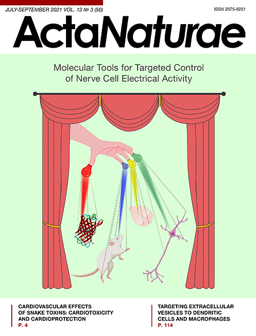
Molecular Tools for Targeted Control of Nerve Cell Electrical Activity. Part I
Abstract
In modern life sciences, the issue of a specific, exogenously directed manipulation of a cell’s biochemistry is a highly topical one. In the case of electrically excitable cells, the aim of the manipulation is to control the cells’ electrical activity, with the result being either excitation with subsequent generation of an action potential or inhibition and suppression of the excitatory currents. The techniques of electrical activity stimulation are of particular significance in tackling the most challenging basic problem: figuring out how the nervous system of higher multicellular organisms functions. At this juncture, when neuroscience is gradually abandoning the reductionist approach in favor of the direct investigation of complex neuronal systems, minimally invasive methods for brain tissue stimulation are becoming the basic element in the toolbox of those involved in the field. In this review, we describe three approaches that are based on the delivery of exogenous, genetically encoded molecules sensitive to external stimuli into the nervous tissue. These approaches include optogenetics (Part I) as well as chemogenetics and thermogenetics (Part II), which are significantly different not only in the nature of the stimuli and structure of the appropriate effector proteins, but also in the details of experimental applications. The latter circumstance is an indication that these are rather complementary than competing techniques.
 52-64
52-64


The p53 Protein Family in the Response of Tumor Cells to Ionizing Radiation: Problem Development
Abstract
Survival mechanisms are activated in tumor cells in response to therapeutic ionizing radiation. This reduces a treatment’s effectiveness. The p53, p63, and p73 proteins belonging to the family of proteins that regulate the numerous pathways of intracellular signal transduction play a key role in the development of radioresistance. This review analyzes the p53-dependent and p53-independent mechanisms involved in overcoming the resistance of tumor cells to radiation exposure.
 65-76
65-76


Genetic Diversity and Evolution of the Biological Features of the Pandemic SARS-CoV-2
Abstract
The new coronavirus infection (COVID-19) represents a challenge for global health. Since the outbreak began, the number of confirmed cases has exceeded 117 million, with more than 2.6 million deaths worldwide. With public health measures aimed at containing the spread of the disease, several countries have faced a crisis in the availability of intensive care units. Currently, a large-scale effort is underway to identify the nucleotide sequences of the SARS-CoV-2 coronavirus that is an etiological agent of COVID-19. Global sequencing of thousands of viral genomes has revealed many common genetic variants, which enables the monitoring of the evolution of SARS-CoV-2 and the tracking of its spread over time. Understanding the current evolution of SARS-CoV-2 is necessary not only for a retrospective analysis of the new coronavirus infection spread, but also for the development of approaches to the therapy and prophylaxis of COVID-19. In this review, we have focused on the general characteristics of SARS-CoV-2 and COVID-19. Also, we have analyzed available publications on the genetic diversity of the virus and the relationship between the diversity and the biological properties of SARS-CoV-2, such as virulence and contagiousness.
 77-89
77-89


Chemiluminescence Detection in the Study of Free-Radical Reactions. Part 1
Abstract
The present review, consisting of two parts, considers the application of the chemiluminescence detection method in evaluating free radical reactions in biological model systems. The first part presents a classification of experimental biological model systems. Evidence favoring the use of chemiluminescence detection in the study of free radical reactions, along with similar methods of registering electromagnetic radiation as electron paramagnetic resonance, spectrophotometry, detection of infrared radiation (IR spectrometry), and chemical methods for assessing the end products of free radical reactions, is shown. Chemiluminescence accompanying free radical reactions involving lipids has been the extensively studied reaction. These reactions are one of the key causes of cell death by either apoptosis (activation of the cytochrome c complex with cardiolipin) or ferroptosis (induced by free ferrous ions). The concept of chemiluminescence quantum yield is also discussed in this article. The second part, which is to be published in the next issue, analyzes the application of chemiluminescence detection using luminescent additives that are called activators, a.k.a. chemiluminescence enhancers, and enhance the emission through the triplet–singlet transfer of electron excitation energy from radical reaction products, followed by light emission with a high quantum yield.
 90-100


Inactivating Gene Expression with Antisense Modified Oligonucleotides
Abstract
Modified nucleotides, including phosphoramidates and mesyl nucleotides, are very effective in inactivating gene expression in bacteria. Gyr A is the target gene in several organisms, including Plasmodium falciparum. Antisense reactions with bacteria infecting citrus plants are promising but incomplete. Human tissue culture cells assayed with a different target are also susceptible to the presence of mesyl oligonucleotides.
 101-105


Research Articles
A Monoiodotyrosine Challenge Test in a Parkinson’s Disease Model
Abstract
Early (preclinical) diagnosis of Parkinson’s disease (PD) is a major challenge in modern neuroscience. The objective of this study was to experimentally evaluate a diagnostic challenge test with monoiodotyrosine (MIT), an endogenous inhibitor of tyrosine hydroxylase. Striatal dopamine was shown to decrease by 34% 2 h after subcutaneous injection of 100 mg/kg MIT to intact mice, with the effect not being amplified by a further increase in the MIT dose. The selected MIT dose caused motor impairment in a neurotoxic mouse model of preclinical PD, but not in the controls. This was because MIT reduced striatal dopamine to the threshold of motor symptoms manifestation only in PD mice. Therefore, using the experimental mouse model of preclinical PD, we have shown that a MIT challenge test may be used to detect latent nigrostriatal dysfunction.
 106-109


A Mouse Model of Nigrostriatal Dopaminergic Axonal Degeneration As a Tool for Testing Neuroprotectors
Abstract
Degeneration of nigrostriatal dopaminergic neurons in Parkinson’s disease begins from the axonal terminals in the striatum and, then, in retrograde fashion, progresses to the cell bodies in the substantia nigra. Investigation of the dynamics of axonal terminal degeneration may help in the identification of new targets for neuroprotective treatment and be used as a tool for testing potential drugs. We have shown that the degeneration rate of dopaminergic axonal terminals changes over time, and that the striatal dopamine concentration is the most sensitive parameter to the action of 1-methyl-4-phenyl-1,2,3,6-tetrahydropyridine (MPTP). This model was validated using neuroprotectors with well-known mechanisms of action: the dopamine transporter inhibitor nomifensine and SEMAX peptide that stimulates the secretion of endogenous neurotrophic factors or acts as an antioxidant. Nomifensine was shown to almost completely protect dopaminergic fibers from the toxic effect of MPTP and maintain the striatal dopamine concentration at the control level. However, SEMAX, slightly but reliably, increased striatal dopamine when administered before MPTP treatment, which indicates that it is more effective as an inductor of endogenous neurotrophic factor secretion rather than as an antioxidant.
 110-113


Targeting Extracellular Vesicles to Dendritic Cells and Macrophages
Abstract
Targeting protein therapeutics to specific cells and tissues is a major challenge in modern medicine. Improving the specificity of protein therapeutic delivery will significantly enhance efficiency in drug development. One of the promising tools for protein delivery is extracellular vesicles (EVs) that are enveloped by a complex lipid bilayer. EVs are secreted by almost all cell types and possess significant advantages: biocompatibility, stability, and the ability to penetrate the blood–brain barrier. Overexpression of the vesicular stomatitis virus protein G (VSV-G) was shown to promote EV formation by the producer cell. We have developed an EV-based system for targeted delivery of protein cargoes to antigen-presenting cells (APCs). In this study, we show that attachment of a recombinant llama nanobody α-CD206 to the N-terminus of a truncated VSV-G increases the selectivity of EV cargo delivery mainly to APCs. These results highlight the outstanding technological and biomedical potential of EV-based delivery systems for correcting the immune response in patients with autoimmune, viral, and oncological diseases.
 114-121


Method for assessment of nucleotide excision repair system efficiency ex vivo
Abstract
The nucleotide excision repair (NER) is one of the main repair systems present in the cells of living organisms. It is responsible for the removal of a wide range of bulky DNA lesions. We succeeded in developing a method for assessing the efficiency of NER in the cell (ex vivo), which is a method based on the recovery of TagRFP fluorescent protein production through repair of the damage that blocks the expression of the appropriate gene. Our constructed plasmids containing bulky nFlu or nAnt lesions near the tagrfp gene promoter were shown to undergo repair in eukaryotic cells (HEK 293T) and that they can be used to analyze the efficiency of NER ex vivo. A comparative analysis of the time dependence of fluorescent cells accumulation after transfection with nFlu- and nAnt-DNA revealed that there are differences in how efficient their repair by the NER system of HEK 293T cells can be. The method can be used to assess the cell repair status and the repair efficiency of different structural damages.
 122-125


 126-128


