


Vol 9, No 3 (2017)
- Year: 2017
- Published: 15.09.2017
- Articles: 13
- URL: https://actanaturae.ru/2075-8251/issue/view/828

Reviews
Virus-Vectored Ebola Vaccines
Abstract
The Ebola virus disease (EVD) is one of the most dangerous infections affecting humans and animals. The first EVD outbreaks occurred in 1976 in Sudan and Zaire. Since then, more than 20 outbreaks have occurred; the largest of which (2014-2016) evolved into an epidemic in West Africa and claimed the lives of more than 11,000 people. Although vaccination is the most effective way to prevent epidemics, there was no licensed vaccine for EVD at the beginning of the latest outbreak. The development of the first vaccines for EVD started in 1980 and has come a long technological way, from inactivated to genetically engineered vaccines based on recombinant viral vectors. This review focuses on virus-vectored Ebola vaccines that have demonstrated the greatest efficacy in preclinical trials and are currently under different phases of clinical trial. Particular attention is paid to the mechanisms of immune response development, which are important for protection from EVD, and the key vaccine parameters necessary for inducing long-term protective immunity against EVD.
 4-11
4-11


Non-bulky Lesions in Human DNA: The Ways of Formation, Repair, and Replication
Abstract
DNA damage is a major cause of replication interruption, mutations, and cell death. DNA damage is removed by several types of repair processes. The involvement of specialized DNA polymerases in replication provides an important mechanism that helps tolerate persistent DNA damage. Specialized DNA polymerases incorporate nucleotides opposite lesions with high efficiency but demonstrate low accuracy of DNA synthesis. In this review, we summarize the types and mechanisms of formation and repair of non-bulky DNA lesions, and we provide an overview of the role of specialized DNA polymerases in translesion DNA synthesis.
12-26
Antitumor Vaccines Based on Dendritic Cells: From Experiments using Animal Tumor Models to Clinical Trials
Abstract
The routine methods used to treat oncological diseases have a number of drawbacks, including non-specific action and severe side effects for patients. Furthermore, tumor diseases are associated with a suppression of the immune system that often leads to the inefficiency of standard treatment methods. The development of novel immunotherapeutic approaches having specific antitumor action and that activate the immune system is of crucial importance. Vaccines based on dendritic cells (DCs) loaded with tumor antigens ex vivo that can activate antitumor cytotoxic T-cell responses stand out among different antitumor immunotherapeutic approaches. This review is focused on analyzing different methods of DC-based vaccine preparation and current research in antitumor DC-based vaccines using animal tumor models and in clinical trials.
27-38
Proteasomes in Protein Homeostasis of Pluripotent Stem Cells
Abstract
Embryonic stem cells (ESCs) and induced pluripotent stem cells (iPSCs) are subjects of high interest not only in basic research, but also in various applied fields, particularly, in regenerative medicine. Despite the tremendous interest to these cells, the molecular mechanisms that control protein homeostasis in these cells remain largely unknown. The ubiquitin-proteasome system (UPS) acts via post-translational protein modifications and protein degradation and, therefore, is involved in the control of virtually all cellular processes: cell cycle, self-renewal, signal transduction, transcription, translation, oxidative stress, immune response, apoptosis, etc. Therefore, studying the biological role and action mechanisms of the UPS in pluripotent cells will help to better understand the biology of cells, as well as to develop novel approaches for regenerative medicine.
39-47
Predicting the Evolutionary Variability of the Influenza A Virus
Abstract
The influenza A virus remains one of the most common and dangerous human health concerns due to its rapid evolutionary dynamics. Since the evolutionary changes of influenza A viruses can be traced in real time, the last decade has seen a surge in research on influenza A viruses due to an increase in experimental data (selection of escape mutants followed by examination of their phenotypic characteristics and generation of viruses with desired mutations using reverse genetics). Moreover, the advances in our understanding are also attributable to the development of new computational methods based on a phylogenetic analysis of influenza virus strains and mathematical (integro-differential equations, statistical methods, probability-theory-based methods) and simulation modeling. Continuously evolving highly pathogenic influenza A viruses are a serious health concern which necessitates a coupling of theoretical and experimental approaches to predict the evolutionary trends of the influenza A virus, with a focus on the H5 subtype.
48-54
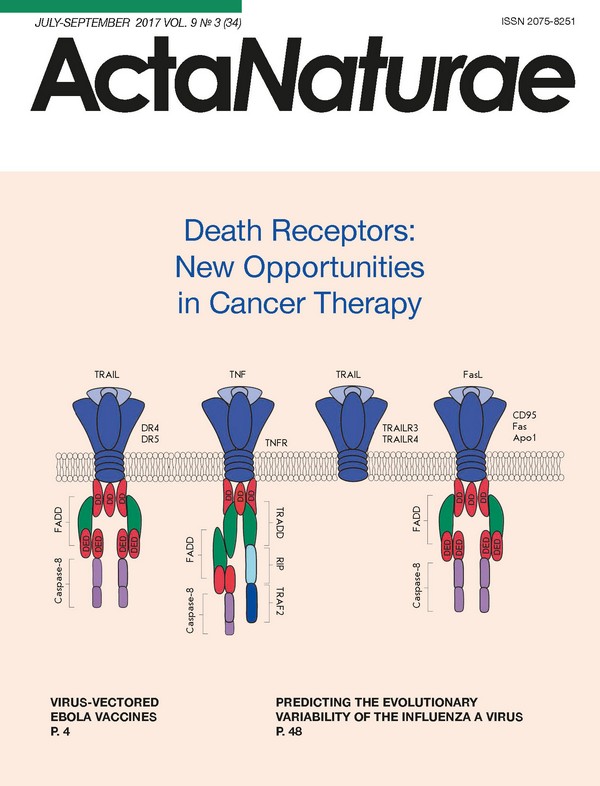
Death Receptors: New Opportunities in Cancer Therapy
Abstract
This article offers a detailed review of the current approaches to anticancer therapy that target the death receptors of malignant cells. Here, we provide a comprehensive overview of the structure and function of death receptors and their ligands, describe the current and latest trends in the development of death receptor agonists, and perform their comparative analysis. In addition, we discuss the DR4 and DR5 agonistic antibodies that are being evaluated at various stages of clinical trials. Finally, we conclude by stating that death receptor agonists may be improved through increasing their stability, solubility, and elimination half-life, as well as by overcoming the resistance of tumor cells. What’s more, effective application of these antibodies requires a more detailed study of their use in combination with other anticancer agents.
55-63
Research Articles
Studying the Specific Activity of the Amide Form of HLDF-6 Peptide using the Transgenic Model of Alzheimer’s Disease
Abstract
The neuroprotective and nootropic activities of the amide form (AF) of the HLDF-6 peptide (TGENHR-NH2 ) were studied in transgenic mice of the B6C3-Tg(APPswe,PSEN1de9)85Dbo (Tg+) line (the animal model of familial Alzheimer’s disease (AD)). The study was performed in 4 mouse groups: group 1 (study group): Tg+ mice intranasally injected with the peptide at a dose of 250 µg/kg; group 2 (active control): Tg+ mice intranasally injected with normal saline; group 3 (control 1): Tg- mice; and group 4 (control 2): C57Bl/6 mice. The cognitive functions were evaluated using three tests: the novel object recognition test, the conditioned passive avoidance task, and the Morris water maze. The results testify to the fact that the pharmaceutical substance (PhS) based on the AF of HLDF-6 peptide at a dose of 250 µg/kg administered intranasally efficiently restores the disturbed cognitive functions in transgenic mice. These results are fully consistent with the data obtained in animal models of Alzheimer’s disease induced by the injection of the beta-amyloid (βA) fragment 25-35 into the giant-cell nucleus basalis of Meynert or by co-injection of the βA fragment 25-35 and ibotenic acid into the hippocampus, and the model of ischemia stroke (chronic bilateral occlusion of carotids, 2VO). According to the overall results, PhS based on AF HLDF-6 was chosen as an object for further investigation; the dose of 250 µg/kg was used as an effective therapeutic dose. Intranasal administration was the route for delivery.
64-70
Change in the Content of Immunoproteasomes and Macrophages in Rat Liver At the Induction of DonorSpecific Tolerance
Abstract
Induction of donor specific tolerance (DST) by the introduction of donor cells into a recipient’s portal vein is one of the approaches used to solve the problem of transplant engraftment. However, the mechanism of DST development remains unclear to this moment. In the present work, we first studied the change in the content of immunoproteasomes and macrophages of the liver at early stages of the development of allospecific portal tolerance in rats by Western blotting and flow cytofluorimetry. On the basis of the data obtained, we can conclude that the induction of DST is an active process characterized by two phases during which the level of the proteasome immune subunits LMP2 and LMP7 in liver mononuclear cells, including Kupffer cells, and the number of Kupffer cells change. The first phase lasts up to 5 days after the beginning of DST induction; the second phase - from 5 to 14 days. In both phases, the level of the subunits LMP2 and LMP7 in the total pool of mononuclear cells and Kupffer cells increases, with maximum values on days 1 and 7. In addition, the total number of Kupffer cells increases in both phases with a shift in several days. The most noticeable changes take place in the second phase. The third day is characterized by a lower content of mononuclear cells expressing immunoproteasomes compared to the control value in native animals. Presumably, at this time point a “window of opportunity” appears for subsequent filling of an empty niche with cells of different subpopulations and, depending on this fact, the development of tolerance or rejection. The results obtained raise the new tasks of finding ways to influence the cellular composition in the liver and the expression of immunoproteasomes on the third day after the beginning of DST induction to block the development of rejection.
71-80
The Spatial Organization of the Intranuclear Structures of Human Brain Dopaminergic Neurons
Abstract
We studied the intranuclear localization of protein nucleophosmin (B23) and ubiquitin in the dopaminergic neurons of human substantia nigra (n = 6, age of 25-87 years) using immunohistochemistry and confocal laser microscopy. Intranuclear ubiquitin-immunopositive bodies that morphologically correspond to Marinesco bodies were found to be present in substantia nigra dopaminergic (tyrosine hydroxylase-immunopositive) neurons but absent in non-dopaminergic neurons. The number of bodies varied from 0 to 6 per cell nucleus. Nucleophosmin (B23) was found in the neuronal nucleolus, with the nucleolus size being constant in the nigral neurons of each individual brain. All the observed neurons had only one large nucleolus with intense nucleophosmin immunoreactivity and a lightly stained region (1-2 µm in diameter) that apparently represents the giant fibrillar center (GFC). An intensely immunostained nucleophosmin-containing granule was often observed at the GFC periphery. Double labeling demonstrated that nucleophosmin-immunoreactive nucleolus and ubiquitin-immunoreactive Marinesco bodies can occur both closely to and remotely from each other. Three-dimensional reconstruction indicates that rounded Marinesco bodies are polymorphic and often have a complex shape, with some flattening and concavities, which may be associated with contact not only with the nucleolus, but also, presumably, with other intranuclear structures free of ubiquitin or nucleophosmin. Ubiquitin-immunoreactive structures with a relatively small size (up to 1 µm in length) and various clastosome-like shapes (Lafarga et al., 2002) often occur near Marinesco bodies. There were no cases of detection of ubiquitin in the nucleoli of dopaminergic neurons and nucleophosmin/B23 in typical Marinesco bodies. The obtained information may be helpful in unraveling the molecular mechanisms of the selective vulnerability of substantia nigra dopaminergic neurons to damaging factors.
81-88
Induction of ICAM-1 Expression in Mouse Embryonic Fibroblasts Cultured on Fibroin-Gelatin Scaffolds
Abstract
Culturing of allogeneic or autologous cells in three-dimensional bioresorbable scaffolds is an important step in the engineering of constructs for regenerative medicine, as well as for experimental systems to study the mechanisms of cell differentiation and cell-to-cell interaction. Artificial substrates can modulate the phenotype and functional activity of immobilized cells. Investigating these changes is important for understanding the fundamental processes underlying cellular interactions in a 3D microenvironment and for improving tissue-engineered structures. In this study, we investigated the expression of the ICAM-1 adhesion molecule in mouse embryonic fibroblasts (MEF) when cultured on gelatin-fibroin scaffolds. Increased expression of ICAM-1 in MEF was detected only under 3D culture conditions both at the mRNA and protein levels. At the same time, the MEF cultured on various substrates did not oerexpress MAdCAM-1, indicating the selective effect of 3D culture conditions on ICAM-1 expression. One possible mechanism for ICAM-1 induction in MEF is associated with the activation of AP-1, since expression of c-Fos and Junb (but not cJun and Jund) was increased in MEF in 3D. When cultured under 2D conditions, the expression level of AP-1 components did not change.
89-93
The Minor Variant of the SingleNucleotide Polymorphism rs3753381 Affects the Activity of a SLAMF1 Enhancer
Abstract
The SLAMF1 gene encodes CD150, a transmembrane glycoprotein expressed on the surface of T and B-lymphocytes, NK-cells, dendritic cells, and subpopulations of macrophages and basophils. We investigated the functional regulatory polymorphisms of the SLAMF1 locus associated with autoimmune processes, using bioinformatics and a mutational analysis of the regulatory elements overlapping with polymorphic positions. In the reporter gene assay in MP-1 and Raji B-cell lines, the enhancer activity of the regulatory region of the locus containing the rs3753381 polymorphism demonstrated a twofold increase upon the introduction of the rs3753381 minor variant (G → A) associated with myasthenia gravis. An analysis of the nucleotide context in the vicinity of rs3753381 revealed that the minor version of this polymorphism improves several binding sites for the transcription factors of FOX and NFAT, and RXR nuclear receptors. All mutations that disrupt any of these sites lead to a decrease in the enhancer activity both in МР-1 and in Raji cells, and each of the two B-cell lines expresses a specific set of these factors. Thus, the minor variant of the rs3753381 polymorphism may contribute to the development of myasthenia gravis by modulating SLAMF1 expression, presumably in pathogenic B-lymphocytes.
94-102
The Effect of the Targeted Recombinant Toxin DARPin-PE40 on the Dynamics of HER2-Positive Tumor Growth
Abstract
The development of targeted toxins based on non-immunoglobulin targeting molecules appears to be one of the most advanced approaches in the targeted therapy of malignant tumors with a high expression of the HER2 receptor. Earlier, we showed that the targeted toxin DARPin-PE40 consisting of the HER2-specific non-immunoglobulin polypeptide (the targeting module) and a fragment of Pseudomonas exotoxin A (the toxic module) exhibits an antitumor effect in vivo against the HER2-positive adenocarcinoma xenograft. In this work, an in-depth analysis of the effect of DARPin-PE40 on the growth dynamics of experimental xenograft tumors was carried out. DARPin-PE40 was shown to inhibit tumor growth at a dose of 25 and 50 µg/animal and to cause tumor node reduction at a dose of 80 µg/animal, followed by growth resumption at the end of therapy. An evaluation of the tumor growth dynamics revealed statistically significant differences in tumor volume in mice in the experimental groups compared to the control group. The results testify to the potential of using the created targeted toxin as an agent for the targeted therapy of HER2-overexpressing tumors.
103-107
Expression Levels of the Uridine-Cytidine Kinase Like-1 Protein As a Novel Prognostic Factor for Hepatitis C VirusAssociated Hepatocellular Carcinomas
Abstract
The expression levels of the two novel oncoproteins uridine-cytidine kinase like-1 (UCKL-1) and mitochondrial ribosomal protein S18-2 (MRPS18-2) were assessed in samples of hepatitis C virus (HCV)-associated hepatocellular carcinoma (HCC) using immunohistochemistry. Tissue microarray (TMA) paraffin blocks were prepared from 42 HCC tumor samples with the corresponding peri-tumor tissues and from 11 tissues of a liver with HCV-induced cirrhosis. We found that the UCKL-1 signal in the liver tissues of the peri-tumor zone in the HCC samples was stronger than that in cirrhosis (50 ± 49.44 vs. 24.27 ± 14.53; p = 0.014). The MRPS18-2 expression was weak, and there was no differences between the groups (p = 0.26). Noteworthy, the UCKL-1 protein was expressed at higher levels in peri-tumor tissues in the cases of HCC recurrence; this was confirmed for 27 older patients (63.78 ± 9.22 vs. 53.53 ± 4.07 years, p < 0.001), in parallel with enhanced UCKL-1 staining in former HCC nodules (62.69 ± 50.4 vs. 26.0 ± 30.19, p = 0.006) and microvascular invasion (p = 0.02). A multivariate analysis of prognostic factors for HCC recurrence showed that the best predictive factors for these conditions were UCKL-1 expression in tumor, vascular invasion, and HCC treatment modality, other than liver transplantation (odds ratios: 1.029, 18.143 and 11.984, R² = 0.633, p = 0.002). In conclusion, the high UCKL-1 expression might be a prognostic factor for HCC relapse, in combination with age and microvascular invasion. MRPS18-2 protein expression has no prognostic significance in the cases of HCV-associated HCC.
108-114