


Vol 6, No 4 (2014)
- Year: 2014
- Published: 15.12.2014
- Articles: 12
- URL: https://actanaturae.ru/2075-8251/issue/view/839

Reviews
Mechanisms of Activation of Voltage-Gated Potassium Channels
Abstract
Voltage-gated potassium ion channels (Kv) play an important role in a variety of cellular processes, including the functioning of excitable cells, regulation of apoptosis, cell growth and differentiation, the release of neurotransmitters and hormones, maintenance of cardiac activity, etc. Failure in the functioning of Kv channels leads to severe genetic disorders and the development of tumors, including malignant ones. Understanding the mechanisms underlying Kv channels functioning is a key factor in determining the cause of the diseases associated with mutations in the channels, and in the search for new drugs. The mechanism of activation of the channels is a topic of ongoing debate, and a consensus on the issue has not yet been reached. This review discusses the key stages in studying the mechanisms of functioning of Kv channels and describes the basic models of their activation known to date.
 10-26
10-26


Research Articles
Regulation of the Target Protein (Transgene) Expression in the Adenovirus Vector Using Agonists of Toll-Like Receptors
Abstract
Replication-defective adenoviral vectors are effective molecular tools for both gene therapy and gene vaccination. Using such vectors one can deliver and express target genes in different epithelial, liver, hematopoietic and immune system cells of animal and human origin. The success of gene therapy and gene vaccination depends on the production intensity of the target protein encoded by the transgene. In this work, we studied influence of Toll-like receptors (TLR) agonists on transduction and expression efficacy of adenoviral vectors in animal and human antigen-presenting cells. We found that agonists of TLR2, 4, 5, 7, 8 and 9 significantly enhance a production of the target protein in cells transduced with adenoviral vector having the target gene insert. The enhancement was observed in dendritic cells and macrophages expressing cytoplasmic (GFP), membrane (HA) or secretory (SEAP) proteins encoded by the respective rAd-vectors. Experiments in mice showed that enhancement of the transgene expression can be achieved in the organism of animals using a pharmaceutical-grade TLR4-agonist. In contrast to other TLR-agonists, the agonist of TLR3 substantially suppressed the expression of transgene in cells transduced with adenoviral vectors having insert of GFP or SEAP target genes. We propose that the enhancement of transgene expression is linked to the activation of MyD88→ NF-kB, while the inhibition of transgene expression depends on TRIF→ IRF signaling pathways. Both of these pathways jointly exploited by TLR4-agonists lead to the enhancement of transgene expression due to the dominant role of the MyD88→ NF-kB signaling.
27-39
STIM1 Protein Activates Store-Operated Calcium Channels in Cellular Model of Huntington’s Disease
Abstract
We have shown that the expression of full-length mutated huntingtin in human neuroblastoma cells (SK-N-SH) leads to an abnormal increase in calcium entry through store-operated channels. In this paper, the expression of the N-terminal fragment of mutated huntingtin (Htt138Q-1exon) is shown to be enough to provide an actual model for Huntington’s disease. We have shown that Htt138Q-1exon expression causes increased store-operated calcium entry, which is mediated by at least two types of channels in SK-N-SH cells with different reversal potentials. Calcium sensor, STIM1, is required for activation of store-operated calcium entry in these cells. The results provide grounds for considering the proteins responsible for the activation and maintenance of the store-operated calcium entry as promising targets for developing novel therapeutics for neurodegenerative diseases.
40-47
Specific Visualization of Tumor Cells Using Upconversion Nanophosphors
Abstract
The development of targeted constructs on the basis of photoluminescent nanoparticles with a high photo- and chemical stability and absorption/emission spectra in the “transparency window” of biological tissues is an important focus area of present-day medical diagnostics. In this work, a targeted two-component construct on the basis of upconversion nanophosphors (UCNPs) and anti-tumor 4D5 scFv was developed for selective labeling of tumor cells overexpressing the HER2 tumor marker characteristic of a number of human malignant tumors. A high affinity barnase : barstar (Bn : Bs) protein pair, which exhibits high stability in a wide range of pH and temperatures, was exploited as a molecular adapter providing self-assembly of the two-component construct. High selectivity for the binding of the two-component 4D5 scFv-Bn : UCNP-Bs construct to human breast adenocarcinoma SK-BR-3 cells overexpressing HER2 was demonstrated. This approach provides an opportunity to produce similar constructs for the visualization of different specific markers in pathogenic tissues, including malignant tumors.
48-53
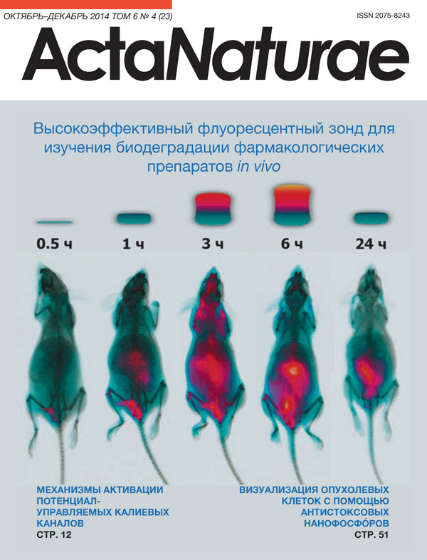
Excessive Labeling Technique Provides a Highly Sensitive Fluorescent Probe for Real-time Monitoring of Biodegradation of Biopolymer Pharmaceuticals in vivo
Abstract
Recombinant proteins represent a large sector of the biopharma market. Determination of the main elimination pathways raises the opportunities to significantly increase their half-lives in vivo. However, evaluation of biodegradation of pharmaceutical biopolymers performed in the course of pre-clinical studies is frequently complicated. Noninvasive pharmacokinetic and biodistribution studies in living organism are possible using proteins conjugated with near-infrared dyes. In the present study we designed a highly efficient probe based on fluorescent dye self-quenching for monitoring of in vivo biodegradation of recombinant human butyrylcholinesterase. The maximum enhancement of integral fluorescence in response to degradation of an intravenously administered enzyme was observed 6 h after injection. Importantly, excessive butyrylcholinesterase labeling with fluorescent dye results in significant changes in the pharmacokinetic properties of the obtained conjugate. This fact must be taken into consideration during future pharmacokinetic studies using in vivo bioimaging.
54-59
Human SLURP-1 and SLURP-2 Proteins Acting on Nicotinic Acetylcholine Receptors Reduce Proliferation of Human Colorectal Adenocarcinoma HT-29 Cells
Abstract
Human secreted Ly-6/uPAR related proteins (SLURP-1 and SLURP-2) are produced by various cells, including the epithelium and immune system. These proteins act as autocrine/paracrine hormones regulating the growth and differentiation of keratinocytes and are also involved in the control of inflammation and malignant cell transformation. These effects are assumed to be mediated by the interactions of SLURP-1 and SLURP-2 with the α7 and α3β2 subtypes of nicotinic acetylcholine receptors (nAChRs), respectively. Available knowledge about the molecular mechanism underling the SLURP-1 and SLURP-2 effects is very limited. SLURP-2 remains one of the most poorly studied proteins of the Ly-6/uPAR family. In this study, we designed for the first time a bacterial system for SLURP-2 expression and a protocol for refolding of the protein from cytoplasmic inclusion bodies. Milligram quantities of recombinant SLURP-2 and its 13С-15N-labeled analog were obtained. The recombinant protein was characterized by NMR spectroscopy, and a structural model was developed. A comparative study of the SLURP-1 and SLURP-2 effects on the epithelial cell growth was conducted using human colorectal adenocarcinoma HT-29 cells, which express only α7-nAChRs. A pronounced antiproliferative effect of both proteins was observed. Incubation of cells with 1 μM SLURP-1 and 1 μM SLURP-2 during 48 h led to a reduction in the cell number down to ~ 54 and 63% relative to the control, respectively. Fluorescent microscopy did not reveal either apoptotic or necrotic cell death. An analysis of the dose-response curve revealed the concentration-dependent mode of the SLURP-1 and SLURP-2 action with EC50 ~ 0.1 and 0.2 nM, respectively. These findings suggest that the α7-nAChR is the main receptor responsible for the antiproliferative effect of SLURP proteins in epithelial cells.
60-66
Investigation of Channel-Forming Activity of Polyene Macrolide Antibiotics in Planar Lipid Bilayers in the Presence of Dipole Modifiers
Abstract
The role of membrane components, sterols, phospholipids and sphingolipids in the formation and functioning of ion-permeable nanopores formed by antifungal macrolide antibiotics, amphotericin B, nystatin and filipin in planar lipid bilayers was studied. Dipole modifiers, flavonoids and styryl dyes, were used as a tool to study the molecular mechanisms of polyene channel-forming activity. The introduction of dipole modifiers into the membrane bathing solutions was shown to change the conductance of single channels and the steadystate transmembrane current induced by polyene antibiotics in the sterol-containing phospholipid-bilayers. The conductance of single amphotericin B channels was found to depend on the dipole potential of the membrane. The experiments with various phospholipids, sterols, and polyenes led to the assumption that the shape of a phospholipid molecule, the presence of double bonds at the positions 7 and 22 of a sterol molecule, the number of conjugated double bonds, and the presence of an amino sugar in the polyene antibiotic molecule are important factors impacting the stability of polyene-lipid complexes forming ion-permeable pores. Experimental and literature data presented in the paper suggest that the channel-forming activity of polyene antibiotics is also affected by the physicochemical properties of polyene-enriched ordered membrane domains.
67-79
A Cytofluorometric Study of Membrane Rafts in Human Monocyte Subsets in Atherosclerosis
Abstract
The peripheral blood monocytes of atherosclerotic patients are pre-activated and have some of the features of tissue macrophages. Their adhesion to the endothelium is 1.5 times higher than that of monocytes from healthy subjects, and they express a number of receptors and antigens typical of tissue macrophages. Additionally, earlier we showed that the biosynthesis of gangliosides, whose main function is the formation of membrane rafts, is significantly activated in blood monocytes from atherosclerotic patients, as well as during the in vitro differentiation of normal monocytes into macrophages. In this study, we investigated the expression of membrane rafts on various monocyte subsets from healthy subjects and atherosclerotic patients. Based on flow cytometry results, the monocytes in the examined atherosclerotic patients were found to differ from those in healthy subjects by a twofold increase in the proportion of the intermediate subset (CD14 ++/CD16 +) and by enhancement in the expression of the fractalkine receptor CX3CR1 on the intermediate and non-classical subsets (CD14 ++/CD16 + and CD14 +/CD16 ++) (2.3 and 1.8 times, respectively). This suggests a pre-activated state of monocytes in atherosclerotic patients. At the same time, the expression of the membrane raft marker on the monocyte subsets was similar in both studied groups. However, a study of the in vitro differentiation of monocytes into macrophages showed that the membrane raft expression increased 2 times as early as on the 1st day of culturing and 3 times on the 7th day compared to that in freshly isolated monocytes. Therefore, it is suggested that monocytes in atherosclerosis accumulate gangliosides that are used to form membrane rafts during the macrophage differentiation after the migration of monocytes into the arterial intima.
80-88
Hct-A Is a New Actinoporin Family from the Heteractis Crispa Sea Anemone
Abstract
Several new actinoporin isoforms with molecular weights of 18995.5 to 19398.7 Da exhibiting a high hemolytic activity were isolated from the tropical sea anemone Heteractis crispa using a combination of liquid chromatography techniques. The actinoporins were demonstrated to occur as mono-, di-, and trimers in aqueous solutions. The sequences of the genes encoding actinoporins were identified, and the amino acid sequences of the new polypeptides belonging to the Hct-A actinoporin family were obtained. The new acinoporins differ in their isoelectric points, the number and localization of charged amino acid residues at the functionally important N-terminal fragment of the molecule, as well as in the charge of a tetrapeptide (amino acid residues 74-77) involved in an electrostatic interaction with the cytoplasmic membrane. A recombinant actinoporin, rHct-A2, with a molecular weight of 19141 Da, pI of 9.64, and hemolytic activity of 4.0 × 10⁴ HU/mg, was obtained. The conductivity of the ion channels formed by rHct-A2 in the BLM was demonstrated to be similar to that of the native actinoporin from H. crispa. The obtained data expand knowledge on the structural and functional relationships of actinoporins and contribute to our understanding of the functioning mechanism of these molecules, which is the basis for the development of compounds with a high biomedical potential. Currently, they are considered as models for obtaining antitumor, antibacterial, and cardiac-stimulating agents.
89-98
Acipensins - Novel Antimicrobial Peptides from Leukocytes of the Russian Sturgeon Acipenser gueldenstaedtii
Abstract
Antimicrobial peptides (AMPs) play an important role in the innate defense mechanisms in humans and animals. We have isolated and studied a set of antimicrobial peptides from leukocytes of the Russian sturgeon Acipenser gueldenstaedtii belonging to a subclass of chondrosteans, an ancient group of bony fish. Structural analysis of the isolated peptides, designated as acipensins (Ac), revealed in leukocytes of the Russian sturgeon six novel peptides with molecular masses of 5336.2 Da, 3803.0 Da, 5173.0 Da, 4777.5 Da, 5449.4 Da, and 2740.2 Da, designated as Ac1-Ac6, respectively. Complete primary structures of all the isolated peptides were determined, and the biological activities of three major components - Ac1, Ac2, and Ac6 - were examined. The peptides Ас1, Ас2, Ас3, Ас4, and Ac5 were found to be the N-terminal acetylated fragments 1-50, 1-35, 1-49, 1-44, and 1-51 of the histone Н2А, respectively, while Ас6 was shown to be the 62-85 fragment of the histone Н2А. The peptides Ac1 and Ac2 displayed potent antimicrobial activity towards Gram-negative and Gram-positive bacteria (Escherichia coli ML35p, Listeria monocytogenes EGD, MRSA ATCC 33591) and the fungus Candida albicans 820, while Ac6 proved effective only against Gram-negative bacteria. The efficacy of Ac 1 and Ac2 towards the fungus and MRSA was reduced upon an increase in the ionic strength of the solution. Ac1, Ac2, and Ac6, at concentrations close to their minimum inhibitory concentrations, enhanced the permeability of the E.coli ML35p outer membrane to the chromogenic marker, but they did not affect appreciably the permeability of the bacterial inner membrane in comparison with a potent pore-forming peptide, protegrin 1. Ac1, Ac2, and Ac6 revealed no hemolytic activity against human erythrocytes at concentrations of 1 to 40 μM and had no cytotoxic effect (1 to 20 μM) on K-562 and U-937 cells in vitro. Our findings suggest that histone-derived peptides serve as important anti-infective host defense molecules.
99-109
The Mechanism of Choline-Mediated Inhibition of Acetylcholine Release in Mouse Motor Synapses
Abstract
The mechanism of action of tonically applied choline, the agonist of α7 nicotinic acetylcholine receptors (nAChRs), to the spontaneous and evoked release of a neurotransmitter in mouse motor synapses in diaphragm neuromuscular preparations using intracellular microelectrode recordings of miniature endplate potentials (MEPPs) and evoked endplate potentials (EPPs) was studied. Exogenous choline was shown to exhibit a presynaptic inhibitory effect on the amplitude and quantal content of EPPs for the activity of neuromuscular junction evoked by single and rhythmic stimuli. This effect was inhibited either by antagonists of α7-nAChRs, such as methyllycaconitine and α-cobratoxin, or by blocking SK-type calcium-activated potassium (K Ca) channels with apamin or blocking intraterminal ryanodine receptors with ryanodine. A hypothesis was put forward that choline in mouse motoneuron nerve terminals can activate presynaptic α7-nAChRs, followed by the release of the stored calcium through ryanodine receptors and activation of SK-type KCa channels, resulting in sustained decay of the quantal content of the evoked neurotransmitter release.
110-115
Short communications
Phosphoryl Guanidines: A New Type of Nucleic Acid Analogues
Abstract
A new type of nucleic acid analogues with a phosphoryl guanidine group is described. Oxidation of polymer-supported dinucleoside 2-cyanoethyl phosphite by iodine in the presence of 1,1,3,3-tetramethyl guanidine yields a dinucleotide with an internucleoside tetramethyl phosphoryl guanidine (Tmg) group as the main product. The Tmg group is stable under conditions of solid-phase DNA synthesis and subsequent cleavage and deprotection with ammonia. Oligonucleotides with one or more Tmg groups bind their complementary DNA or RNA with affinity similar to that of natural oligodeoxyribonucleotides.
116-118