Vol 10, No 1 (2018)
- Year: 2018
- Published: 15.03.2018
- Articles: 9
- URL: https://actanaturae.ru/2075-8251/issue/view/826
Reviews
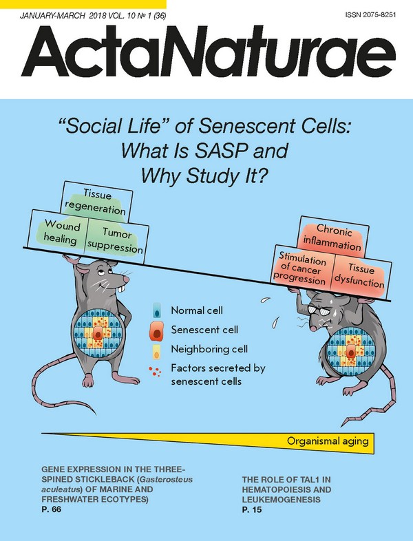
“Social Life” of Senescent Cells: What Is SASP and Why Study It?
Abstract
Cellular senescence was first described as a failure of normal human cells to divide indefinitely in culture. Until recently, the emphasis in the study of cell senescence has been focused on the accompanying intracellular processes. The focus of the attention has been on the irreversible growth arrest and two important physiological functions that rely on it: suppression of carcinogenesis due to the proliferation loss of damaged cells, and the acceleration of organism aging due to the deterioration of the tissue repair mechanism with age. However, the advances of the past years have revealed that senescent cells can impact the surrounding tissue microenvironment, and, thus, that the main consequences of senescence are not solely mediated by intracellular alterations. Recent studies have provided evidence that a pool of molecules secreted by senescent cells, including cytokines, chemokines, proteases and growth factors, termed the senescence-associated secretory phenotype (SASP), via autocrine/paracrine pathways can affect neighboring cells. Today it is clear that SASP functionally links cell senescence to various biological processes, such as tissue regeneration and remodeling, embryonic development, inflammation, and tumorigenesis. The present article aims to describe the “social” life of senescent cells: basically, SASP constitution, molecular mechanisms of its regulation, and its functional role.
 4-14
4-14


The Role of TAL1 in Hematopoiesis and Leukemogenesis
Abstract
TAL1 (SCL/TAL1, T-cell acute leukemia protein 1) is a transcription factor that is involved in the process of hematopoiesis and leukemogenesis. It participates in blood cell formation, forms mesoderm in early embryogenesis, and regulates hematopoiesis in adult organisms. TAL1 is essential in maintaining the multipotency of hematopoietic stem cells (HSC) and keeping them in quiescence (stage G0). TAL1 forms complexes with various transcription factors, regulating hematopoiesis (E2A/HEB, GATA1-3, LMO1-2, Ldb1, ETO2, RUNX1, ERG, FLI1). In these complexes, TAL1 regulates normal myeloid differentiation, controls the proliferation of erythroid progenitors, and determines the choice of the direction of HSC differentiation. The transcription factors TAL1, E2A, GATA1 (or GATA2), LMO2, and Ldb1 are the major components of the SCL complex. In addition to normal hematopoiesis, this complex may also be involved in the process of blood cell malignant transformation. Upregulation of C-KIT expression is one of the main roles played by the SCL complex. Today, TAL1 and its partners are considered promising therapeutic targets in the treatment of T-cell acute lymphoblastic leukemia.
 15-23
15-23


Pathogenesis of Type 1 Diabetes Mellitus and Rodent Experimental Models
Abstract
The global prevalence of diabetes mellitus and its severe complications is on the rise. The study of the pathogenesis of the onset and the progression of complications related to the disease, as well as the search for new therapeutic agents and methods of treatment, remains relevant. Experimental models are extremely important in the study of diabetes. This survey contains a synthesis of the most commonly used experimental animal models described in scientific literature. The mechanisms of the streptozotocin model are also analyzed and discussed, as it is considered as the most adequate and easily reproducible diabetes model. A review of the significant advantages and disadvantages of the described models has also been conducted.
 24-33
24-33


Research Articles
The Effect of TNF and VEGF on the Properties of Ea.hy926 Endothelial Cells in a Model of Multi-Cellular Spheroids
Abstract
Endothelial cells play a major role in the development of inflammation and neoangiogenesis in cancer and chronic inflammatory diseases. In 3D cultures, cells are under conditions that closely resemble those existing in healthy and disease-stricken human organs and tissues. Therefore, the development of a 3D model based on the Ea.hy926 endothelial cell line is an urgent need in molecular and cellular biology. Cell cultivation on an anti-adhesive substrate under static conditions was shown to lead to the formation of spheroids (3D cultures). Expression of ICAM-1 and VEGFR-2 and production of cytokines were screened in 2D and 3D cultures in the presence of TNF and VEGF. According to flow cytometry and confocal microscopy data, TNF significantly increased the expression of the cell adhesion molecule ICAM-1 in both 2D and 3D cultures but did not affect the expression level of VEGFR-2. Increased production of pro-inflammatory (IL-8, IL-6, IP-10) and anti-inflammatory (IL-10, TGF-β 1-3) factors was observed in spontaneous 3D cultures but not in 2D cultures, which was confirmed by flow cytometry and qPCR. TNF-induced secretion of IL-10, GM-CSF, and IL-6 was 11-, 4.7-, and 1.6-fold higher, respectively, in 3D cultures compared to 2D cultures. Thus, the use of a Ea.hy926 3D cell culture is a promising approach in studying the effects of anti- and pro-inflammatory agents on endothelial cells.
 34-42
34-42


The PTENP1 Pseudogene, Unlike the PTEN Gene, Is Methylated in Normal Endometrium, As Well As in Endometrial Hyperplasias and Carcinomas in Middle-Aged and Elderly Females
Abstract
The tumor suppressor PTEN controls multiple cellular functions, including cell cycle, apoptosis, senescence, transcription, and mRNA translation of numerous genes. In tumor cells, PTEN is frequently inactivated by genetic mutations and epimutations.rolex yacht master replica The aim of this study was to investigate the methylation patterns of the PTEN gene and its pseudogene PTENP1 as potential genetic markers of endometrial hyperplasia (EH) and endometrial carcinoma (EC). Methylation of the 5’-terminal regions of the PTEN and PTENP1 sequences was studied using methyl-sensitive PCR of genomic DNA isolated from 57 cancer, rolex datejust special edition replica43 endometrial hyperplasia, and normal tissue samples of 24 females aged 17-34 years and 19 females aged 45-65 years, as well as 20 peripheral venous blood samples of EC patients. None of the analyzed DNA samples carried a methylated PTEN gene. On the contrary, the PTENP1 pseudogene was methylated in all analyzed tissues, except for the peripheral blood. Comparison of PTENP1 methylation rates revealed no differences between the EC and EH groups (0.80 < p < 0.50). In all these groups, the methylation level was high (71-77% in patients vs. 58% in controls). Differences in PTENP1 methylation rates between normal endometrium in young (4%) and middle-aged and elderly (58%) females were significant (p < 0.001). These findings suggest that PTENP1 pseudogene methylation may reflect age-related changes in the body and is not directly related to the endometrium pathology under study. It is assumed that,real rolex submariner vs fake depending on the influence of a methylated PTENP1 pseudogene on PTEN gene expression, the pseudogene methylation may protect against the development of EC and/or serve as a marker of a precancerous condition of endometrial cells.
 43-50
43-50


A Highly Productive CHO Cell Line Secreting Human Blood Clotting Factor IX
Abstract
Hemophilia B patients suffer from an inherited blood-clotting defect and require regular administration of blood-clotting factor IX replacement therapy. Recombinant human factor IX produced in cultured CHO cells is nearly identical to natural, plasma-derived factor IX and is widely used in clinical practice. Development of a biosimilar recombinant human factor IX for medical applications requires the generation of a clonal cell line with the highest specific productivity possible and a high level of specific procoagulant activity of the secreted factor IX. We previously developed plasmid vectors, p1.1 and p1.2, based on the untranslated regions of the translation elongation factor 1 alpha gene from Chinese hamster. These vectors allow one to perform the methotrexate- driven amplification of the genome-integrated target genes and co-transfect auxiliary genes linked to various resistance markers. The natural open reading frame region of the factor IX gene was cloned in the p1.1 vector plasmid and transfected to CHO DG44 cells. Three consecutive amplification rounds and subsequent cell cloning yielded a producer cell line with a specific productivity of 10.7 ± 0.4 pg/cell/day. The procoagulant activity of the secreted factor IX was restored nearly completely by co-transfection of the producer cells by p1.2 plasmids bearing genes of the soluble truncated variant of human PACE/furin signal protease and vitamin K oxidoreductase from Chinese hamster. The resulting clonal cell line 3B12-86 was able to secrete factor IX in a protein-free medium up to a 6 IU/ml titer under plain batch culturing conditions. The copy number of the genome- integrated factor IX gene for the 3B12-86 cell line was only 20 copies/genome; the copy numbers of the genome-integrated genes of PACE/furin and vitamin K oxidoreductase were 3 and 2 copies/genome, respectively. Factor IX protein secreted by the 3B12-86 cell line was purified by three consecutive chromatography rounds to a specific activity of up to 230 IU/mg, with the overall yield > 30%. The developed clonal producer cell line and the purification process employed in this work allow for economically sound industrial-scale production of biosimilar factor IX for hemophilia B therapy.
 51-65
51-65


Gene Expression in the Three-Spined Stickleback (Gasterosteus aculeatus) of Marine and Freshwater Ecotypes
Abstract
Three-spine stickleback (Gasterosteus aculeatus) is a well-known model organism that is routinely used to explore microevolution processes and speciation, and the number of studies related to this fish has been growing recently. The main reason for the increased interest is the processes of freshwater adaptation taking place in natural populations of this species. Freshwater three-spined stickleback populations form when marine water three-spined sticklebacks fish start spending their entire lifecycle in freshwater lakes and streams. To boot, these freshwater populations acquire novel biological traits during their adaptation to a freshwater environment. The processes taking place in these populations are of great interest to evolutionary biologists. Here, we present differential gene expression profiling in G. aculeatus gills, which was performed in marine and freshwater populations of sticklebacks. In total, 2,982 differentially expressed genes between marine and freshwater populations were discovered. We assumed that differentially expressed genes were distributed not randomly along stickleback chromosomes and that they are regularly observed in the “divergence islands” that are responsible for stickleback freshwater adaptation.
 66-74
66-74


New Experimental Models of Retinal Degeneration for Screening Molecular Photochromic Ion Channel Blockers
Abstract
Application of molecular photochromic ion channel blockers to recover the visual function of a degenerated retina is one of the promising trends in photopharmacology. To this day, several photochromic azobenzene-based compounds have been proposed and their functionality has been demonstrated on cell lines and knockout mouse models. Further advance necessitates testing of the physiological activity of a great number of new compounds. The goal of this study is to propose animal models of photoreceptor degeneration that are easier to obtain than knockout mouse models but include the main features required for testing the physiological activity of molecular photoswitches. Two amphibian-based models were proposed. The first model was obtained by mechanical deletion of the photoreceptor outer segments. The second model was obtained by intraocular injection of tunicamycin to induce the degeneration of rods and cones. To test our models, we used 2-[(4-{(E)-[4-(acryloylaminophenyl]diazenyl}phenyl)amino]-N,N,N-triethyl-2-oxoethanammonium chloride (AAQ), one of the compounds that have been studied in other physiological models. The electroretinograms recorded from our models before and after AAQ treatment are in agreement with the results obtained on knockout mouse models and reported in other studies. Hence, the proposed models can be used for primary screening of molecular photochromic ion channel blockers.
 75-84
75-84


Influence of the Linking Order of Fragments of HA2 and M2e of the influenza A Virus to Flagellin on the Properties of Recombinant Proteins
Abstract
The ectodomain of the M2 protein (M2e) and the conserved fragment of the second subunit of hemagglutinin (HA2) are promising candidates for broadly protective vaccines. In this paper, we report on the design of chimeric constructs with differing orders of linkage of four tandem copies of M2e and the conserved fragment of HA2 (76-130) from phylogenetic group II influenza A viruses to the C-terminus of flagellin. The 3D-structure of two chimeric proteins showed that interior location of the M2e tandem copies (Flg-4M2e-HA2) provides partial α-helix formation nontypical of native M2e on the virion surface. The C-terminal position of the M2e tandem copies (Flg-HA2-4M2e) largely retained its native M2e conformation. These conformational differences in the structure of the two chimeric proteins were shown to affect their immunogenic properties. Different antibody levels induced by the chimeric proteins were detected. The protein Flg-HA2-4M2e was more immunogenic as compared to Flg-4M2e-HA2, with the former offering full protection to mice against a lethal challenge. We obtained evidence suggesting that the order of linkage of target antigens in a fusion protein may influence the 3D conformation of the chimeric construct, which leads to changes in immunogenicity and protective potency.
 85-94
85-94


