


Рецепторы смерти: новые возможности в терапии онкологических заболеваний
- Авторы: Украинская В.M.1, Степанов A.В.1,2, Глаголева И.С.2, Кнорре В.Д.1, Белогуров A.A.1,2, Габибов A.Г.1,2
-
Учреждения:
- Институт биоорганической химии им. академиков М.М. Шемякина и Ю.А. Овчинникова РАН
- Казанский (Приволжский) федеральный университет
- Выпуск: Том 9, № 3 (2017)
- Страницы: 55-63
- Раздел: Обзоры
- Дата подачи: 17.01.2020
- Дата публикации: 15.09.2017
- URL: https://actanaturae.ru/2075-8251/article/view/10376
- DOI: https://doi.org/10.32607/20758251-2017-9-3-55-63
- ID: 10376
Цитировать

Аннотация
В представленном обзоре рассмотрены подходы к терапии опухолей с использованием клеточных рецепторов смерти. Описано строение и функционирование рецепторов смерти и их лигандов. Охарактеризованы направления разработки агонистов рецепторов смерти и проведен их сравнительный анализ. Также рассмотрены антитела - агонисты рецепторов DR4 и DR5, находящиеся на различных этапах клинических исследований. Сделан вывод, что усовершенствование агонистов рецепторов смерти предполагает повышение их стабильности, растворимости и периода полувыведения, а также преодоление резистентности опухолевых клеток. Для эффективного применения антител требуется более подробное изучение их комбинаций с другими противоопухолевыми агентами.
Ключевые слова
Полный текст
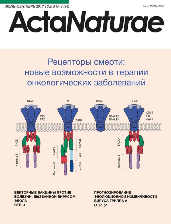
ВВЕДЕНИЕ Основными методами терапии онкологических за болеваний являются химиотерапия в комбинации с хирургическим вмешательством. В силу отсутствия селективности по отношению к злокачественным клеткам эта стратегия приводит к разнообразным побочным эффектам и возможным осложнениям. Поэтому привлекательной выглядит идея создания специфичных препаратов, направленно подавляю щих развитие раковых клеток, таких, как антитела и их производные [1]. Важным этапом в разработке противоопухолевых препаратов стало создание ини циирующих апоптоз агентов. Известно, что при зло качественной трансформации часто нарушаются пути активации апоптоза - физиологического про цесса, регулирующего количество клеток и играю щего ключевую роль в элиминации поврежденных, ненужных или зараженных клеток [2]. Все более глу бокое понимание механизмов регуляции программи руемой гибели клеток привело к появлению новых агентов, способных перезапустить апоптоз в злока чественных клетках. Основную долю современных терапевтических агентов, инициирующих апоптоз, составляют низкомолекулярные вещества, которые часто вызывают развитие системных осложнений [3]. Принципиально другой мишенью противоопухоле вой терапии является поиск агонистов суперсемейства рецепторов фактора некроза опухоли (tumor necrosis factor receptor superfamily, TNFRSF). Отдельную группу в данном суперсемействе составляют так на зываемые рецепторы смерти, содержащие домен смерти (death domain). К рецепторам смерти относят ся рецептор фактора некроза опухоли 1 (TNFR1), ре цептор фактора некроза опухоли 6 (CD95, FasR, Apo1), рецептор смерти 4 (DR4), рецептор смерти 5 (DR5) и др. Наиболее перспективными мишенями для тар гетной терапии опухолевых заболеваний считаются DR4 и DR5, так как их экспрессия в раковых клетках повышена по сравнению с нормальными клетками [4, 5]. В здоровых клетках механизмы апоптоза регули руются группой антиапоптотических белков, к приме ру, FLICE-подобным ингибирующим белком (с-FLIP), подавляющим активацию каспазы-8. Также в клетке присутствуют белки семейства Bcl-2, которые, обра зуя гетерокомплекс с каспазами, подавляют актив ность апоптотического сигнала [6, 7]. СТРУКТУРА РЕЦЕПТОРОВ СМЕРТИ 4 И 5 DR4 и DR5 - трансмембранные белки типа I - состо ят из трех доменов: внеклеточного, трансмембран ного и внутриклеточного. Внутриклеточный домен содержит гомологичную цитоплазматическую по следовательность - домен смерти. DR5 может су ществовать в виде двух изоформ DR5(L) и DR5(S): в короткой форме между цистеиновыми последова тельностями и трансмембранным участком отсут ствуют 29 аминокислотных остатков, что, однако, никак не сказывается на его функциональной актив ности [8]. Рецепторы DR4 и DR5 присутствуют во многих клетках человека, включая тимус, печень, лейкоци ты крови, активированные Т-лимфоциты, тонкий ки шечник. Эти рецепторы обнаружены в некоторых ли ниях опухолевых клеток, таких, как Jurkat [9], Ramos [10], HeLa [11], Colo205 [12] и др. Домены смерти DR4 и DR5 идентичны на 64%, а обогащенные цистеином домены - на 66% [13]. Взаимодействие рецептора с лигандом TRAIL (Tumor necrosis factor ligand superfamily number 10) сначала происходит в N-концевой части внеклеточ ного домена, когда лиганд соединяется с первым ци стеиновым доменом, так называемым участком пред варительного взаимодействия (preligand assembly domain, PLAD) [14]. Эта последовательность не при нимает непосредственного участия в олигомериза ции рецептора, но позволяет стабилизировать поло жение лиганда относительно рецептора [15]. Ранее обнаружили, что тримеризация лиганда происходит в присутствии иона Zn+2 [16], который нековалентно связывается с цистеин-богатыми доменами TRAIL. При стабилизации TRAIL в мономере рецептора происходят конформационное изменение, затем передвижение в липидных рафтах мембраны и об разование активной формы тримера [17]. В инициа ции апоптоза ключевую роль играет домен смерти, к которому за счет гомотипического взаимодействия домена смерти адаптерного белка с доменом смер ти рецептора (DD-DD) присоединяется адаптерный белок. Такими адаптерными молекулами являются белок FADD (Fas-associated DD-protein), взаимодей ствующий с доменом смерти Fas-рецептора, и белок TRADD (TNFR1-associated DD-protein), взаимодей ствующий с доменом смерти TNFR1-рецептора [18]. TRADD и FADD также включают дополнительные модули белок-белкового взаимодействия, называе мые эффекторными доменами (DED) [19]. Эти моду ли принимают участие в присоединении прокаспазы 8/10 и регуляторного белка c-FLIP. Мультибелковый комплекс, образующийся между доменом смерти ре цептора, FADD и каспазами 8/10, называется сиг нальным комплексом, индуцирующим смерть (death inducing signaling complex, DISC) [20] (рис. 1). После формирования DISC апоптотический сигнал переда ется на инициаторные каспазы. АКТИВАЦИЯ АПОПТОЗА Апоптоз является сложным энергозависимым про цессом, основанным на каскаде молекулярных пре вращений. На данный момент известны два пути раз вития апоптоза: рецепторный и митохондриальный. После формирования DISC апоптотический сиг нал передается на инициаторные каспазы. Каспазы существуют в клетке в форме неактивных прокаспаз (32-56 кДа), представляющих собой мономеры, со стоящие из N-концевого домена, большой (17-21 кДа) и малой (10-13 кДа) субъединиц и коротких связу ющих областей [21]. Существует несколько теорий, описывающих процесс активации каспаз. Согласно одной - кластеризация каспаз на комплексе DISC приводит к их самоактивации за счет аутокатали тического процессинга. Согласно другой - сборка инициаторных каспаз благоприятствует их димери зации, в результате чего N-концевой продомен и свя зующие области в составе каждого из мономеров рас щепляются, при этом большая и малая субъединица формируют гетеродимеры [22]. Субстратная специфичность инициаторных каспаз ограничивается эффекторными каспазами и проа поптотическим белком Bid [23]. При активации ини циаторных каспаз 8/10 в составе комплекса DISC происходит дальнейшая активация эффекторных каспаз-3 и -7, обладающих ферментативной актив ностью. Сайтом гидролиза эффекторных каспаз яв ляется остаток Asp в тетрапептидном мотиве [24, 25]. Активация эффекторных каспаз приводит к запу ску множества сигнальных путей, контролирующих жизнедеятельность клетки. Митохондриальный путь апоптоза чаще всего активируется внутриклеточными факторами в от вет на различные сигналы: разрушение ДНК, появ ление активных форм кислорода, накопление бел ка с нарушенным фолдингом и др. Данный процесс регулируется белками семейства Bcl-2, в которое входит фактор Bid, активируемый путем протеоли за под действием каспазы-8 [26]. В активированной форме tBid вызывает пермеабилизацию митохон дриальной мембраны, выход цитохрома с и фор мирование апоптосомы, вызывающей активацию инициаторной каспазы-9 [27]. Это ключевой момент в развитии митохондриального пути апоптоза, далее происходит активация эффекторных каспаз (рис. 2). Оба пути, и рецепторный, и митохондриальный, приводят к активации цитоплазматических эндону клеаз, деградирующих ДНК, и протеаз, разрушаю щих внутриклеточные белки. Сами каспазы-3, -7 и -6 расщепляют цитокератин и плазматическую мем брану, что обуславливает морфологические изме нения, которые происходят в любой апоптотической клетке [28]. TRAIL TRAIL, как и фактор некроза опухоли (TNF), входит в суперсемейство фактора некроза опухоли (TNFSF) и участвует в регуляции жизненно важных биологи ческих функций у позвоночных [29]. TRAIL содер жит в своей структуре два антипараллельных бета складчатых листа, которые формируют структуру бета-сендвича [30]. Обладающий единственным ци стеиновым остатком, TRAIL способен образовывать хелатные комплексы с цинком. Субъединицы взаи модействуют друг с другом по схеме голова-к-хвосту с образованием пирамиды, усеченной к хвосту гомо тримера [31]. Также TRAIL содержит значительное количество ароматических остатков, восемь из ко торых находятся на поверхности внутреннего листа и создают гидрофобную платформу для взаимодей ствия с соседними субъединицами. При создании терапевтических конструкций TRAIL имеет ряд преимуществ перед другими ли гандами, вызывающими апоптоз. Основной его осо бенностью является отсутствие цитотоксичности в отношении нормальных клеток, в отличие от Fas лиганда и TNF. Предполагается, что это обусловле но специфичностью TRAIL к расположенным на по верхности здоровых клеток рецепторам-«ловушкам» DcR1 (Decoy receptor 1) и DcR2 (Decoy receptor 2), у которых отсутствует внутриклеточный домен смерти [32]. Рецепторы-«ловушки» ингибируют раз витие апоптоза, конкурируя с DR4 и DR5 за связы вание с TRAIL. Также сам рецептор DcR2 может связываться с DR4, образуя лиганднезависимый комплекс [33]. Тем не менее, не до конца ясно, что еще обеспечивает выживаемость здоровых клеток, так как рецепторы-«ловушки» обнаруживают также на опухолевых клетках, чувствительных к TRAIL. РЕЗИСТЕНТНОСТЬ ОПУХОЛЕВЫХ КЛЕТОК К TRAIL Существуют различные причины возникновения резистентности к TRAIL. Не последнюю роль в этом играют репрессоры сигнала апоптоза: к которым относятся белок с-FLIP, белки ингибиторы (IAP), транскрипционный фактор NF-kB и др. [34]. Чрезмерная экспрессия антиапоптотических белков, относящихся к семейству Bcl-2, может вно сить свой вклад в развитие резистентности к TRAIL в различных видах опухолевых клеток [35]. Доказано, что присутствие «разрезанной» формы с-FLIP в со ставе комплекса DISC препятствует активации ка спазы-8 [36]. Причиной резистентности к TRAIL мо гут быть различные мутации в белках, участвующих в сигнальном пути апоптоза. Так, мутация проапоп тотического белка Bax приводит к устойчивости эпи телиальных клеток рака толстой кишки [37]. К примеру, TRAIL-чувствительные линии ней роэктодермальных опухолевых клеток (PNET) экс прессируют в необходимом количестве мРНК и белок каспазы-8, в отличие от TRAIL-резистентных клеток PNET, что вызвано метилированием гена, кодирую щего каспазу. Показано, что в клетках PNET рези стентность к TRAIL сохраняется даже при сверхэк спрессии рецепторов к TRAIL [38, 39]. Высокий уровень транскрипционного фактора NFkB в опухолевых клетках может вызывать не только повышенную экспрессию рецепторов DR4 и DR5 [40], но и развитие резистентности к TRAIL, что вызвано повышенным синтезом антиапоптотических белков, которые регулируются данным фактором [41]. Описанные выше варианты покрывают далеко не все пути развития резистентности в опухолевых клетках. Ее преодоление является основным направ лением в разработке новых агентов, активирующих рецепторы DR4 и DR5. АГОНИСТЫ TRAIL-R В ТЕРАПИИ РАКА На данный момент разработано множество страте гий направленного воздействия на TRAIL-R - это различные формы рекомбинантного растворимого TRAIL человека (Apo2L или Dulanermin), антитела агонисты DR4, DR5 и др. [42]. Эти агенты безопасны и хорошо переносятся пациентами [43, 44]. Активность идеального терапевтического аген та, действующего на TRAIL-зависимый апоптоз, должна соответствовать природному лиганду, по добно антителу с высокой аффинностью связывать ся с рецептором, и его период полувыведения дол жен обеспечивать продолжительную персистенцию в кровотоке. Рекомбинантный TRAIL человека ак тивирует оба рецептора смерти, но его применение лимитируется быстрым расщеплением в крови и ма лым временем полувыведения. Также TRAIL может связываться с рецепторами-«ловушками», способ ными подавлять активацию апоптоза [45]. В качестве альтернативы TRAIL созданы антитела, взаимодействующие только с рецепторами смерти и не влияю щими на рецепторы-«ловушки». Они относительно безопасны, обладают улучшенными фармакокине тическими свойствами по сравнению с рекомбинант ным TRAIL, но специфичны только к одному виду рецептора. Несмотря на существующие ограничения, на этапе клинических испытаний находится много различных агентов, воздействующих на рецепторы смерти как в виде монотерапии, так и в комплексном подходе. На данный момент разработаны инновационные стратегии, направленные на усовершенствование структуры TRAIL-лиганда. Первая рекомбинантная версия TRAIL содержала TNF-гомологичный домен, на N-концевой участок которого добавлен полиги стидиновый кластер [46] или FLAG-эпитоп [47]. Эти фрагменты позволяют улучшить процесс очистки белка. Несмотря на то что два модифицированных белка показали эффективность in vitro и in vivo, их использование затрудняет появление гепатотоксич ности. Для увеличения стабильности комплекса TRAIL создано несколько его модификаций. Один из под ходов состоит в присоединении к TRAIL мотива лей циновой (LZ-TRAIL) или изолейциновой застежки (iz-TRAIL). Аналогичным решением является объ единение TRAIL с тенасцином-С для стабилизации и олигомеризации молекулы. Эти агенты показали большую, чем у Dulanermin, активность in vivo и in vitro и безопасность в отношении гепатоцитов [48]. Совсем недавно несколько исследовательских групп разработали новый принцип стабилизации TRAIL, основанный на одноцепочечном TRAIL (scTRAIL) [49]. При данном подходе молекула экс прессируется в виде тримера, в котором три доме на объединены линкером по типу голова-к-хвосту. Правильно собранная конструкция исключает воз можность ошибки при ее экспрессии и предупреж дает неспецифические взаимодействия с другими молекулами. Это дает scTRAIL преимущества перед его аналогами и обеспечивает эффективность в отно шении некоторых резистентных опухолевых линий. Для увеличения периода полувыведения TRAIL к нему присоединяют молекулы с лучшей фармако кинетикой, такие, как сывороточный альбумин чело века или полиэтиленгликоль (ПЭГ). По результатам in vivo исследований пэгилирование iz-TRAIL уве личило период полувыведения, стабильность и рас творимость молекулы [50]. АНТИТЕЛА Антитела к TRAIL-R1 (Mapatumumab [51]) и TRAIL-R2 (Conatumumab [52], Lexatumumab [53], Tigatuzumab [54] и Drozitumab [55]) показали опре деленную эффективность в доклинических исследо ваниях. При проведении клинических исследований все антитела были безопасными и более стабильными по сравнению с TRAIL. Антитела, эффективные на I стадии клинических исследований, на II стадии ис пользовали как в качестве монотерапии, так и в ком бинации с цисплатином, паклитакселом [56] и други ми противоопухолевыми средствами. В качестве монотерапии эффективными оказались антитела Mapatumumab и Сonatumumab. При при менении Mapatumumab клинические улучшения наблюдались у 14 из 17 пациентов с неходжкинской лимфомой. У 29% пациентов с немелкоклеточным ра ком легких и у 32% с колоректальным раком наблю далась продолжительная ремиссия [57, 58]. Комбинация Сonatumumab с паклитакселом и карбоплатином в качестве впервые назначенной терапии при немелкоклеточном раке легкого ока залась более эффективной, чем при применении только паклитаксела и карбоплатина [59], в отличие от Mapatumumab, эффективность которого в анало гичной комбинации не подтвердилась [60]. Также Сonatumumab был эффективен в комби нации со стандартной химиотерапией FOLFIRI и га нитумабом в качестве вторичной терапии колорек тального рака - выявлено повышение выживаемости пациентов, находящихся в состоянии ремиссии [61]. Tigatuzumab (CS-1008) в комбинации с гентамици ном показал хорошую переносимость при метастази рующем раке печени. Доля пациентов с объективным ответом составила 13.1% [62]. Dulanermin - рекомбинантный аналог лиганда ре цепторов смерти, был проверен на пациентах с опу холями различного происхождения. На стадии до клинических исследований этот препарат проявил активность против хондробластомы, колоректального рака и других опухолей. Но, к сожалению, клиниче ские исследования не подтвердили данные доклини ческих испытаний [63]. Таким образом, для эффективного применения агонистов рецепторов смерти требуется индиви дуальный подход к каждому пациенту, так как су ществует вероятность резистентности опухолевых клеток к данной терапии. Одним из принципов ее преодоления может быть поиск определенных био маркеров резистентности, которые помогли бы оха рактеризовать клетки с высоким уровнем экспрессии рецепторов смерти, потенциально чувствительных к воздействию антител [64]. Одним из подходов преодоления резистентности является применение генетически модифициро ванных Т-лимфоцитов. На основе одноцепочечного антитела к DR4, слитого с химерным антигенным рецептором Т-клеток (chimeric antigen receptor, CAR), получены Т-клетки, специфически элимини рующие опухолевые клетки с DR4. Было показано, что при взаимодействии с опухолевыми клетками модифицированные химерным рецептором Т-клетки запускают не только индуцированный DR4 путь апоптоза, но и механизмы заложенного цитотокси ческого действия Т-клеток [65, 66]. ПЕПТИДНЫЕ АГОНИСТЫ РЕЦЕПТОРОВ СМЕРТИ Перспективным представляется поиск подходящих пептидов, агонистов DR4 и DR5. К преимуществам пептидов относится их способность селективно свя зываться только с определенным рецептором смерти [67]. Для отбора пептидных лигандов используется технология фагового дисплея, которая, связывая ге нотип с фенотипом, выбирает пептиды с агонистиче скими свойствами. Эти пептиды могут связываться с рецепторами DR4 и DR5 и активировать их. Методом фагового дисплея был отобран пептид YCKVILTHRCY, который специфично связывается с DR5. Для увеличения растворимости на концы пеп тида добавили остатки Tyr. Изучены свойства этого пептида в мономерной и димерной (два ковалентно соединенных мономера) форме. Установлено, что обе формы взаимодействуют с DR5 и вызывают развитие апоптоза в клеточной линии рака кишечника Colo205. Эффективность мономера может быть связана с тем, что в высоких концентрациях пептид может агреги ровать в водной среде, так как содержит много ги дрофобных групп [68]. Аналогичным способом был отобран пептид GRVCLTLCSRLT, проявляющий вы сокую аффинность к DR5 (IC50 = 30 нM). Показано, что ключевую роль во взаимодействии с рецептором играет аминокислотная последовательность LTL [69]. ЗАКЛЮЧЕНИЕ Разработаны различные подходы к воздействию на опухолевые клетки, в том числе и через пути апоптоза. К сожалению, многие из них оказываются неработоспособными из-за резистентности клеток, неэффективности и неустойчивости самих терапев тических агентов. Другие же открывают новые воз можности терапии опухолевых заболеваний. Более подробное изучение сигнальных путей рецепторов смерти позволит создавать новые агенты, которые смогут направленно воздействовать на раковые клет ки. В свою очередь, для эффективного применения существующих антител к рецепторам смерти требу ется более подробное изучение возможности их при менения в формате сочетанной терапии.
Об авторах
В. M. Украинская
Институт биоорганической химии им. академиков М.М. Шемякина и Ю.А. Овчинникова РАН
Email: stepanov.aleksei.v@gmail.com
Россия
A. В. Степанов
Институт биоорганической химии им. академиков М.М. Шемякина и Ю.А. Овчинникова РАН; Казанский (Приволжский) федеральный университет
Автор, ответственный за переписку.
Email: stepanov.aleksei.v@gmail.com
Россия
И. С. Глаголева
Казанский (Приволжский) федеральный университет
Email: stepanov.aleksei.v@gmail.com
Россия
В. Д. Кнорре
Институт биоорганической химии им. академиков М.М. Шемякина и Ю.А. Овчинникова РАН
Email: stepanov.aleksei.v@gmail.com
Россия
A. A. Белогуров
Институт биоорганической химии им. академиков М.М. Шемякина и Ю.А. Овчинникова РАН; Казанский (Приволжский) федеральный университет
Email: stepanov.aleksei.v@gmail.com
Россия
A. Г. Габибов
Институт биоорганической химии им. академиков М.М. Шемякина и Ю.А. Овчинникова РАН; Казанский (Приволжский) федеральный университет
Email: stepanov.aleksei.v@gmail.com
Россия
Список литературы
- Deyev S.M., Lebedenko E.N., Petrovskaya L.E., Dolgikh D.A., Gabibov A.G., Kirpichnikov M.P. // Russian Chemical Reviews. 2015, V.84, №1, P.1-26
- Wong R.S. // J. Exp. Clin. Cancer Res. 2011, V.30, P.87
- Parameswaran N., Patial S. // Crit. Rev. Eukaryot. Gene Expr. 2010, V.20, №2, P.87-103
- Strater J., Hinz U., Walczak H., Mechtersheimer G., Koretz K., Herfarth C., Moller P., Lehnert T. // Clin. Cancer Res. 2002, V.8, №12, P.3734-3740
- Pan G., O’Rourke K., Chinnaiyan A.M., Gentz R., Ebner R., Ni J., Dixit V.M. // Science. 1997, V.276, №5309, P.111-113
- Chinnaiyan A.M., Dixit V.M. // Semin. Immunol. 1997, V.9, №1, P.69-76
- Reed J.C. // Semin. Hematol. 1997, V.34, №4, P.9-19
- Wang T.T., Jeng J. // Breast Cancer Res. Treat. 2000, V.61, №1, P.87-96
- Natoni A., MacFarlane M., Inoue S., Walewska R., Majid A., Knee D., Stover D.R., Dyer M.J., Cohen G.M. // Br. J. Haematol. 2007, V.139, №4, P.568-577
- MacFarlane M., Kohlhaas S.L., Sutcliffe M.J., Dyer M.J., Cohen G.M. // Cancer Research 2005, V.65, №24, P.11265-11270
- Ren Y.G., Wagner K.W., Knee D.A., Aza-Blanc P., Nasoff M., Deveraux Q.L. // Mol. Biol. Cell. 2004, V.15, №11, P.5064-5074
- Chiron D., Pellat-Deceunynck C., Maillasson M., Bataille R., Jego G. // J. Immunol. 2009, V.183, №7, P.4371-4377
- Pan G., Ni J., Wei Y.F., Yu G., Gentz R., Dixit V.M. // Science. 1997, V.277, №5327, P.815-818
- Clancy L., Mruk K., Archer K., Woelfel M., Mongkolsapaya J., Screaton G., Lenardo M.J., Chan F.K. // Proc. Natl. Acad. Sci. USA. 2005, V.102, №50, P.18099-18104
- Sfikakis P.P., Tsokos G.C. // Clin. Immunol. 2011, V.141, №3, P.231-235
- Ozoren N., El-Deiry W.S. // Semin. Cancer Biol. 2003, V.13, №2, P.135-147
- Marconi M., Ascione B., Ciarlo L., Vona R., Garofalo T., Sorice M., Gianni A.M., Locatelli S.L., Carlo-Stella C., Malorni W. // Cell Death Dis. 2013, V.4, P.e863
- Kuang A.A., Diehl G.E., Zhang J., Winoto A. // J. Biol. Chem. 2000, V.275, №33, P.25065-25068
- Riley J.S., Malik A., Holohan C., Longley D.B. // Cell Death Dis. 2015, V.6, P.e1866
- Kischkel F.C., Lawrence D.A., Chuntharapai A., Schow P., Kim K.J., Ashkenazi A. // Immunity. 2000, V.12, №6, P.611-620
- Earnshaw W.C. // Nature 1999, V.397, №6718, P.387-389
- Riedl S.J., Shi Y. // Nat. Rev. Mol. Cell. Biol. 2004, V.5, №11, P.897-907
- Huang K., Zhang J., O’Neill K.L., Gurumurthy C.B., Quadros R.M., Tu Y., Luo X. // J. Biol. Chem. 2016, V.291, №22, P.11843-11851
- MacKenzie S.H., Clark A.C. // Adv. Exp. Med. Biol. 2012, V.747, P.55-73
- Thornberry N.A. // Br. Med. Bull. 1997, V.53, №3, P.478-490
- Mukae N., Enari M., Sakahira H., Fukuda Y., Inazawa J., Toh H., Nagata S. // Proc. Natl. Acad. Sci. USA. 1998, V.95, №16, P.9123-9128
- Finucane D.M., Bossy-Wetzel E., Waterhouse N.J., Cotter T.G., Green D.R. // J. Biol. Chem. 1999, V.274, №4, P.2225-2233
- Slee E.A., Adrain C., Martin S.J. // J. Biol. Chem. 2001, V.276, №10, P.7320-7326
- Banks T.A., Rickert S., Benedict C.A., Ma L., Ko M., Meier J., Ha W., Schneider K., Granger S.W., Turovskaya O. // J. Immunol. 2005, V.174, №11, P.7217-7225
- Cha S.S., Kim M.S., Choi Y.H., Sung B.J., Shin N.K., Shin H.C., Sung Y.C., Oh B.H. // Immunity. 1999, V.11, №2, P.253-261
- Zakaria A., Picaud F., Guillaume Y.C., Gharbi T., Micheau O., Herlem G. // J. Mol. Recognit. 2016, V.29, №9, P.406-414
- Baritaki S., Huerta-Yepez S., Sakai T., Spandidos D.A., Bonavida B. // Mol. Cancer Ther. 2007, V.6, №4, P.1387-1399
- Marsters S.A., Sheridan J.P., Pitti R.M., Huang A., Skubatch M., Baldwin D., Yuan J., Gurney A., Goddard A.D., Godowski P. // Curr. Biol. 1997, V.7, №12, P.1003-1006
- Prasad S., Kim J.H., Gupta S.C., Aggarwal B.B. // Trends Pharmacol. Sci. 2014, V.35, №10, P.520-536
- Sivaprasad U., Shankar E., Basu A. // Cell Death Differ. 2007, V.14, №4, P.851-860
- Guseva N.V., Rokhlin O.W., Taghiyev A.F., Cohen M.B. // Breast Cancer Res. Treat. 2008, V.107, №3, P.349-357
- LeBlanc H., Lawrence D., Varfolomeev E., Totpal K., Morlan J., Schow P., Fong S., Schwall R., Sinicropi D., Ashkenazi A. // Nat. Med. 2002, V.8, №3, P.274-281
- Kim H.S., Lee J.W., Soung Y.H., Park W.S., Kim S.Y., Lee J.H., Park J.Y., Cho Y.G., Kim C.J., Jeong S.W. // Gastroenterology. 2003, V.125, №3, P.708-715
- Agolini S.F., Shah K., Jaffe J., Newcomb J., Rhodes M., Reed J.F., 3rd. I.O. // J. Trauma. 1997, V.43, №3, P.395-399
- Ravi R., Bedi G.C., Engstrom L.W., Zeng Q., Mookerjee B., Gelinas C., Fuchs E.J., Bedi A. // Nat. Cell. Biol. 2001, V.3, №4, P.409-416
- Kwon H.R., Lee K.W., Dong Z., Lee K.B., Oh S.M. // Biochem. Biophys. Res. Commun. 2010, V.391, №1, P.830-834
- Lemke J., von Karstedt S., Zinngrebe J., Walczak H. // Cell Death Differ. 2014, V.21, №9, P.1350-1364
- Joy A.M., Beaudry C.E., Tran N.L., Ponce F.A., Holz D.R., Demuth T., Berens M.E. // J. Cell Sci. 2003, V.116, P.4409-4417
- Dimberg L.Y., Anderson C.K., Camidge R., Behbakht K., Thorburn A., Ford H.L. // Oncogene. 2013, V.32, №11, P.1341-1350
- Milutinovic S., Kashyap A.K., Yanagi T., Wimer C., Zhou S., O’Neil R., Kurtzman A.L., Faynboym A., Xu L., Hannum C.H. // Mol. Cancer Ther. 2016, V.15, №1, P.114-124
- Pitti R.M., Marsters S.A., Ruppert S., Donahue C.J., Moore A., Ashkenazi A. // J. Biol. Chem. 1996, V.271, №22, P.12687-12690
- Wiley S.R., Schooley K., Smolak P.J., Din W.S., Huang C.P., Nicholl J.K., Sutherland G.R., Smith T.D., Rauch C., Smith C.A. // Immunity. 1995, V.3, №6, P.673-682
- Rozanov D.V., Savinov A.Y., Golubkov V.S., Rozanova O.L., Postnova T.I., Sergienko E.A., Vasile S., Aleshin A.E., Rega M.F., Pellecchia M. // Mol. Cancer Ther. 2009, V.8, №6, P.1515-1525
- Siegemund M., Pollak N., Seifert O., Wahl K., Hanak K., Vogel A., Nussler A.K., Gottsch D., Munkel S., Bantel H. // Cell Death Dis. 2012, V.3, P.e295
- Harris J.M., Chess R.B. // Nat. Rev. Drug Discov. 2003, V.2, №3, P.214-221
- Greco F.A., Bonomi P., Crawford J., Kelly K., Oh Y., Halpern W., Lo L., Gallant G., Klein J. // Lung Cancer. 2008, V.61, №1, P.82-90
- Doi T., Murakami H., Ohtsu A., Fuse N., Yoshino T., Yamamoto N., Boku N., Onozawa Y., Hsu C.P., Gorski K.S. // Cancer Chemother. Pharmacol. 2011, V.68, №3, P.733-741
- Merchant M.S., Geller J.I., Baird K., Chou A.J., Galli S., Charles A., Amaoko M., Rhee E.H., Price A., Wexler L.H. // J. Clin. Oncol. 2012, V.30, №33, P.4141-4147
- Reck M., Krzakowski M., Chmielowska E., Sebastian M., Hadler D., Fox T., Wang Q., Greenberg J., Beckman R.A., von Pawel J. // Lung Cancer. 2013, V.82, №3, P.441-448
- Kang Z., Chen J.J., Yu Y., Li B., Sun S.Y., Zhang B., Cao L. // Clin. Cancer Res. 2011, V.17, №10, P.3181-3192
- Sasaki Y., Nishina T., Yasui H., Goto M., Muro K., Tsuji A., Koizumi W., Toh Y., Hara T., Miyata Y. // Cancer Sci. 2014, V.105, №7, P.812-817
- Younes A., Vose J.M., Zelenetz A.D., Smith M.R., Burris H.A., Ansell S.M., Klein J., Halpern W., Miceli R., Kumm E. // Br. J. Cancer. 2010, V.103, №12, P.1783-1787
- Trarbach T., Moehler M., Heinemann V., Kohne C.H., Przyborek M., Schulz C., Sneller V., Gallant G., Kanzler S. // Br. J. Cancer. 2010, V.102, №3, P.506-512
- Paz-Ares L., Balint B., de Boer R.H., van Meerbeeck J.P., Wierzbicki R., De Souza P., Galimi F., Haddad V., Sabin T., Hei Y.J. // J. Thorac. Oncol. 2013, V.8, №3, P.329-337
- von Pawel J., Harvey J.H., Spigel D.R., Dediu M., Reck M., Cebotaru C.L., Humphreys R.C., Gribbin M.J., Fox N.L., Camidge D.R. // Clin. Lung Cancer. 2014, V.15, №3, P.188-196
- Cohn A.L., Tabernero J., Maurel J., Nowara E., Sastre J., Chuah B.Y., Kopp M.V., Sakaeva D.D., Mitchell E.P., Dubey S. // Ann. Oncol. 2013, V.24, №7, P.1777-1785
- Forero-Torres A., Infante J.R., Waterhouse D., Wong L., Vickers S., Arrowsmith E., He A.R., Hart L., Trent D., Wade J. // Cancer Med. 2013, V.2(6), P.925-932
- Pan Y., Xu R., Peach M., Huang C.P., Branstetter D., Novotny W., Herbst R.S., Eckhardt S.G., Holland P.M. // Br. J. Cancer. 2011, V.105, №12, P.1830-1838
- Dine J.L., O’Sullivan C.C., Voeller D., Greer Y.E., Chavez K.J., Conway C.M., Sinclair S., Stone B., Amiri-Kordestani L., Merchant A.S. // Breast Cancer Res. Treat. 2016, V.155, №2, P.235-251
- Kobayashi E., Kishi H., Ozawa T., Hamana H., Nakagawa H., Jin A., Lin Z., Muraguchi A. // Biochem. Biophys. Res. Commun. 2014, V.453, №4, P.798-803
- Jin A., Ozawa T., Tajiri K., Lin Z., Obata T., Ishida I., Kishi H., Muraguchi A. // Eur. J. Immunol. 2010, V.40, №12, P.3591-3593
- Ladner R.C., Sato A.K., Gorzelany J., de Souza M. // Drug Discov. Today. 2004, V.9, №12, P.525-529
- Vrielink J., Heins M.S., Setroikromo R., Szegezdi E., Mullally M.M., Samali A., Quax W.J. // FEBS J. 2010, V.277, №7, P.1653-1665
- Li B., Russell S.J., Compaan D.M., Totpal K., Marsters S.A., Ashkenazi A., Cochran A.G., Hymowitz S.G., Sidhu S.S. // J. Mol. Biol. 2006, V.361, №3, P.522-536
- Wakelee H.A., Patnaik A., Sikic B.I., Mita M., Fox N.L., Miceli R., Ullrich S.J., Fisher G.A., Tolcher A.W. // Ann. Oncol. 2010, V.21, №2, P.376-381
- Hotte S.J., Hirte H.W., Chen E.X., Siu L.L., Le L.H., Corey A., Iacobucci A., MacLean M., Lo L., Fox N.L. // Clin. Cancer Res. 2008, V.14, №11, P.3450-3455
- Mom C.H., Verweij J., Oldenhuis C.N., Gietema J.A., Fox N.L., Miceli R., Eskens F.A., Loos W.J., de Vries E.G., Sleijfer S. // Clin. Cancer Res. 2009, V.15, №17, P.5584-5590
- Ciuleanu T., Bazin I., Lungulescu D., Miron L., Bondarenko I., Deptala A., Rodriguez-Torres M., Giantonio B., Fox N.L., Wissel P. // Ann. Oncol. 2016, V.27, №4, P.680-687
- Herbst R.S., Kurzrock R., Hong D.S., Valdivieso M., Hsu C.P., Goyal L., Juan G., Hwang Y.C., Wong S., Hill J.S. // Clin. Cancer Res. 2010, V.16, №23, P.5883-5891
- Demetri G.D., Le Cesne A., Chawla S.P., Brodowicz T., Maki R.G., Bach B.A., Smethurst D.P., Bray S., Hei Y.J., Blay J.Y. // Eur. J. Cancer 2012, V.48, №4, P.547-563
- Fuchs C.S., Fakih M., Schwartzberg L., Cohn A.L., Yee L., Dreisbach L., Kozloff M.F., Hei Y.J., Galimi F., Pan Y. // Cancer. 2013, V.119, №24, P.4290-4298
- Kindler H.L., Richards D.A., Garbo L.E., Garon E.B., Stephenson J.J. Jr., Rocha-Lima C.M., Safran H., Chan D., Kocs D.M., Galimi F. // Ann. Oncol. 2012, V.23, №11, P.2834-2842
- Forero-Torres A., Shah J., Wood T., Posey J., Carlisle R., Copigneaux C., Luo F.R., Wojtowicz-Praga S., Percent I., Saleh M. // Cancer Biother. Radiopharm. 2010, V.25, №1, P.13-19
- Forero-Torres A., Varley K.E., Abramson V.G., Li Y., Vaklavas C., Lin N.U., Liu M.C., Rugo H.S., Nanda R., Storniolo A.M. // Clin. Cancer Res. 2015, V.21, №12, P.2722-2729
- Cheng A.L., Kang Y.K., He A.R., Lim H.Y., Ryoo B.Y., Hung C.H., Sheen I.S., Izumi N., Austin T., Wang Q. // J. Hepatol. 2015, V.63, №4, P.896-904
- Ciprotti M., Tebbutt N.C., Lee F.T., Lee S.T., Gan H.K., McKee D.C., O’Keefe G.J., Gong S.J., Chong G., Hopkins W. // J. Clin. Oncol. 2015, V.33, №24, P.2609-2616
- Rocha Lima C.M., Bayraktar S., Flores A.M., MacIntyre J., Montero A., Baranda J.C., Wallmark J., Portera C., Raja R., Stern H. // Cancer Invest. 2012, V.30, №10, P.727-731
- Soria J.C., Smit E., Khayat D., Besse B., Yang X., Hsu C.P., Reese D., Wiezorek J., Blackhall F. // J. Clin. Oncol. 2010, V.28, №9, P.1527-1533
- Soria J.C., Mark Z., Zatloukal P., Szima B., Albert I., Juhasz E., Pujol J.L., Kozielski J., Baker N., Smethurst D. // J. Clin. Oncol. 2011, V.29, №33, P.4442-4451
- Wainberg Z.A., Messersmith W.A., Peddi P.F., Kapp A.V., Ashkenazi A., Royer-Joo S., Portera C.C., Kozloff M.F. // Clin. Colorectal Cancer. 2013, V.12, №4, P.248-254
- Cheah C.Y., Belada D., Fanale M.A., Janikova A., Czucman M.S., Flinn I.W., Kapp A.V., Ashkenazi A., Kelley S., Bray G.L. // Lancet Haematol. 2015, V.2, №4, P.e166-174
- Camidge D.R., Herbst R.S., Gordon M.S., Eckhardt S.G., Kurzrock R., Durbin B., Ing J., Tohnya T.M., Sager J., Ashkenazi A. // Clin. Cancer Res. 2010, V.16, №4, P.1256-1263
Дополнительные файлы

