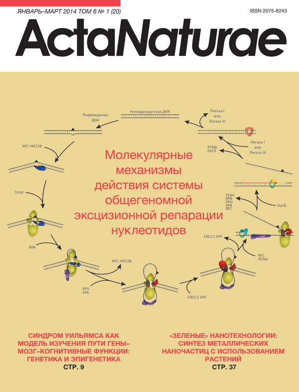
Generation of iPS Cells from Human Hair Follice Dermal Papilla Cells

Cover Page

Cite item

Abstract

Dermal papilla (DP) cells are unique regional stem cells of the skin that induce formation of a hair follicle and its regeneration cycle. DP are multipotent stem cells; therefore we supposed that the efficiency of DPC reprogramming could exceed that of dermal fibroblasts reprogramming. We generated induced pluripotent stem cells from human DP cells using lentiviral transfection with Oct4, Sox2, Klf4, and c-Myc, and cultivation of cells both in a medium supplemented with valproic acid and at a physiological level of oxygen (5%). The efficiency of DP cells reprogramming was ~0.03%, while the efficiency of dermal fibroblast reprogramming under the same conditions was ~0.01%. Therefore, we demonstrated the suitability of DP cells as an alternative source of iPS cells.

About the authors

I. A. Muchkaeva

Koltsov Institute of Developmental Biology, Russian Academy of Sciences; Pirogov Russian National Research Medical University

Author for correspondence.
Email: izomerizaciya@list.ru
Россия

E. B. Dashinimaev

Koltsov Institute of Developmental Biology, Russian Academy of Sciences; Pirogov Russian National Research Medical University

Email: izomerizaciya@list.ru
Россия

A. S. Artyuhov

Pirogov Russian National Research Medical University

Email: izomerizaciya@list.ru
Россия

E. P. Myagkova

Koltsov Institute of Developmental Biology, Russian Academy of Sciences; Pirogov Russian National Research Medical University

Email: izomerizaciya@list.ru
Россия

E. A. Vorotelyak

Koltsov Institute of Developmental Biology, Russian Academy of Sciences; Pirogov Russian National Research Medical University

Email: izomerizaciya@list.ru
Россия

Y. Y. Yegorov

Engelhardt Institute of Molecular Biology, Russian Academy of Sciences

Email: izomerizaciya@list.ru
Россия

K. S. Vishnyakova

Engelhardt Institute of Molecular Biology, Russian Academy of Sciences

Email: izomerizaciya@list.ru
Россия

J. E. Kravchenko

Engelhardt Institute of Molecular Biology, Russian Academy of Sciences; Shemyakin and Ovchinnikov Instituse of Bioorganic Chemistry, Russian Academy of Sciences

Email: izomerizaciya@list.ru
Россия

S. P. Chumakov

Engelhardt Institute of Molecular Biology, Russian Academy of Sciences; Shemyakin and Ovchinnikov Instituse of Bioorganic Chemistry, Russian Academy of Sciences

Email: izomerizaciya@list.ru
Россия

V. V. Terskikh

Koltsov Institute of Developmental Biology, Russian Academy of Sciences

Email: izomerizaciya@list.ru
Россия

A. V. Vasiliev

Koltsov Institute of Developmental Biology, Russian Academy of Sciences

Email: izomerizaciya@list.ru
Россия

References

  1. Oliver R.F. // J. Embryol. Exp. Morphol. 1967. V. 18. № 1. P. 43-51.
  2. Ohuama M., Kobayashi T., Sasaki T., Shimizu A., Amagai M. // J. Cell Sci. 2012. V. 125. № Pt17. P. 4114-4125.
  3. Shutova M.V., A.N. Bogomazova, M.A. Lagarkova, S.L. Kiselev // ActaNaturae. 2009. V. 1. № 2. P. 104-106.
  4. Takahashi K., Yamanaka S. // Cell. 2006. V. 126. № 4. P. 663-676.
  5. Liskovykh M. A., Chuykin I. A., Ranjan A., Safina D. A., Tolkunova E. N., Minina Yu. M., Zhdanova N. S., Dyban P. A., Mullins J., Kostyleva E. I. et al. // Tsitologiya. 2011. V. 53. № 12. P. 927-934.
  6. Higgins C., Ito M., Inoue K., Richardson G., Jahoda K., Cristiano A. // Exp. Dermatol. 2010. V. 19. № 6. P. 568.
  7. Higgins C., Ito M., Inoue K., Richardson G., Jahoda K., Cristiano A. // J. Invest. Dermatol. 2012. V. 136. № 6. P. 1725-1727.
  8. Medvedev S.P., Grigor’eva E.V., Shevchenko A.I., Malakhova A.A., Dementyeva E.V., Shilov A.A., Pokushalov E.A., Zaidman A.M., Aleksandrova M.A., Plotnikov E.Y. et. al. // Stem Cells Dev. 2011. V. 20. № 6. P. 1099-1112.
  9. Warren L., Manos P.D., Ahfeldt T., Loh Y.H., Li H., Lau F., Ebina W., Mandal P.K., Smith Z.D., Meissner A. et al. // Cell Stem Cell. 2010. V. 7. № 5. P. 618-630.
  10. Chermnykh E. S., Radyukhina N. V., Rutkevich P. N., Shevelyov A. Y., Vlasik T. N., Vorotelyak E. A., Vasiliev A. V., Terskikh V. V. // Tsitologiya. V. 52. № 3. P. 219-224.
  11. Ohnuki M., Takahashi K., Yamanaka S. Generation and Characterization of Human Indused Pluripotent Stem Cells. Curr. Protoc. Stem Cell Biol. 2009. Chapter 4:Unit 4A.2. doi: 10.1002/9780470151808.sc04a02s9.
  12. Li R., Liang J., Ni S., Zhou T., Qing X., Li H., He W., Chen J., Li F., Zhuang Q., et. al. // Cell Stem Cell. 2010. 7 № 1. P. 51-63.
  13. Kim D., Kim C.H., Moon J.I., Chung Y.G., Chang M.Y., Han B.S., Ko S., Yang E., Cha K.Y., Lanza R. et. al. // Cell Stem Cell. 2009. V. 4. № 6. P. 472-476.
  14. Marchetto M.C., Yeo G.W., Kainohana O., Marsala M., Gage F.H., Muotri A.R. // PLoS One. 2009. V. 4. № 9. e7076.
  15. Gnedeva K.Y. // Candidate Thesis, M., 2012.
  16. Yehezkel S., Rebibo-Sabbah A., Segev Y., Tzukerman M., Shaked R., Huber I., Gepstein L., Skorecki K., Selig S. // Epigenetics. 2011. V. 6. № 1. P. 63-75.
  17. Rendl M., Polak L., Fuchs E. // Genes Develop.2008. V. 22. № 4. P. 543-557.
  18. Prokhorova, T.A., L.M. Harkness, U. Frandsen, N. Ditzel, H. D. Schroder, J.S. Burns, M. Kassem. // Stem Cells Dev. 2009. V. 18. № 1. P. 47-54.
  19. Zhang W.Y., de Almeida P.E., Wu J.C. // 2012. Teratoma formation: A tool for monitoring pluripotency in stem cell research. StemBook [Internet], doi/10.3824/stembook.1.53.1. http://www.stembook.org.

Supplementary files

Supplementary Files
Action
1. JATS XML

Copyright (c) 2014 Muchkaeva I.A., Dashinimaev E.B., Artyuhov A.S., Myagkova E.P., Vorotelyak E.A., Yegorov Y.Y., Vishnyakova K.S., Kravchenko J.E., Chumakov S.P., Terskikh V.V., Vasiliev A.V.

Creative Commons License
This work is licensed under a Creative Commons Attribution 4.0 International License.

This website uses cookies

You consent to our cookies if you continue to use our website.

About Cookies