


Биомедицинские наносистемы для in vivo детоксикации: от пассивных систем доставки к функциональным наноустройствам и нанороботам
- Авторы: Паширова Т.Н.1, Шайхутдинова З.М.1,2, Миронов В.Ф.1, Массон П.2
-
Учреждения:
- Институт органической и физической химии им. А.Е. Арбузова, ФИЦ Казанский научный центр РАН
- Казанский (Приволжский) федеральный университет
- Выпуск: Том 15, № 1 (2023)
- Страницы: 4-12
- Раздел: Обзоры
- Дата подачи: 15.02.2023
- Дата принятия к публикации: 20.03.2023
- Дата публикации: 03.05.2023
- URL: https://actanaturae.ru/2075-8251/article/view/15681
- DOI: https://doi.org/10.32607/actanaturae.15681
- ID: 15681
Цитировать

Полный текст
СПИСОК СОКРАЩЕНИЙ ЛЭ – липидные эмульсии; RM-PL – эритролипосома; LSPD – перитонеальный диализ с липосомами; ФОС – фосфорорганические соединения; E – фермент; T – токсикант.
ВВЕДЕНИЕ
Долгое время методы профилактики и лечения заболеваний человека основывались исключительно на введении химических или биологических лекарственных препаратов. Начиная с момента открытия в 1964 году первых липосомальных систем, современная стратегия наномедицины направлена на инкапсулирование и стабилизацию низкомолекулярных лекарственных веществ или макромолекул с помощью различных типов наноносителей для преодоления биологических барьеров, повышения биодоступности, снижения нежелательной токсичности для здоровых тканей и адресной доставки [1, 2]. Несмотря на то, что нанотерапевтические препараты уже одобрены для применения в клинике и/или проходят клинические испытания [3, 4], наномедицина по-прежнему сталкивается с низкой эффективностью во многих приложениях, так, например, в среднем только 0.7% цитотоксических препаратов, инкапсулированных в наноносители, достигают солидных опухолевых образований [5]. Начиная с 2008 года наблюдается значительный рост публикаций, описывающих получение нанотерапевтических препаратов нового поколения, так называемых «интеллектуальных наноносителей», модифицированных различными лигандами, обеспечивающими адресную доставку и чувствительность к различным стимулам [6, 7].
На сегодняшний день возникла потребность в альтернативных биомедицинских системах, таких, как роботизированные наноустройства, которые в отличие традиционных пассивных нанотерапевтических препаратов способны выполнять различные биомедицинские функции, включая прецизионную хирургию, биозондирование, детектирование и визуализацию in vivo, адресную доставку лекарств, а в последнее время и детоксикацию [8, 9]. Наноробототехника долгое время была лишь фантастикой, впервые выдвинутой в 1959 году концепцией Ричарда Фейнмана, лауреата Нобелевской премии по физике, о микроскопических механических хирургах, продвигающихся по кровеносному сосуду. Вскоре, в 1966 году, концепция «хирурга» была представлена в научно-фантастическом фильме «Фантастическое путешествие», где миниатюрная подводная лодка использовалась для очистки кровеносного сосуда от тромба. За последние несколько десятилетий область научной фантастики стала реальностью. С использованием различных материалов, технологий и методов управления разработаны наноустройства разнообразной формы и размеров. В качестве наноустройств часто упоминаются микро/наномоторы [10], микро/нанопловцы [11], микро/наномашины [12], микро/нанонасосы [13], микро/наноракеты [14] и т.д. [15].
Существуют следующие определения наноустройств. Наномашины – это наноразмерные механические устройства, способные преобразовывать энергию в точное механическое движение [16]. Микро/нанобиомедицинские устройства – это структуры, которыми можно управлять и приводить в движение в живом организме с помощью химических или биогибридных источников [17]. Это миниатюрные структуры на основе наноматериалов, спроектированные таким образом, чтобы автономно двигаться и эффективно выполнять запрограммированные задачи даже в труднодоступных местах органов/тканей/клеток [18]. Таким образом, роботизированные наноустройства представляют собой инструменты нового поколения, которые могут продвигаться и/или направляться эндогенными и экзогенными стимулами для целенаправленного и персонализированного терапевтического применения. Решение проблем в практических клинических приложениях с использованием нанороботов все еще находится в зачаточном состоянии [19]. К ключевым факторам, которые необходимо учитывать при разработке и успешном применении идеальных биомедицинских терапевтических наноустройств в клинике, относятся:
- биосовместимость с телом пациента;
- способность загрузки/выгрузки лекарств, визуализирующих агентов и т.д.;
- контролируемое движение и возможность отслеживания во времени с помощью методов медицинской визуализации;
- контролируемая деградация без образования каких-либо токсичных метаболитов в теле пациента.
Номенклатура микро/наноустройств основана на их конструкции, геометрии, механизме движения и характере вращения. Как правило, самодвижение наноустройств осуществляется за счет:
а) преобразования энергии химических и ферментативных [20, 21] реакций в механическую [22]. Такие наноустройства двигаются в определенном направлении за счет энергии ферментативных или различных химических реакций [23, 24], например, i) наноустройства, передвигающиеся за счет образования пузырьков газа (водорода, кислорода и т.д.); ii) самоэлектрофоретические наноустройства, работающие по принципу разницы окислительно-восстановительного потенциала; iii) самодиффузионные наноустройства, где механизм движения осуществляется за счет градиента концентрации при образовании продуктов реакции;
б) влияния внешних стимулов [25] (магнитное, акустическое, световое поле), т.е. это стимул-чувствительные наноустройства;
в) биологические/биогибридные наноустройства, где движение обусловлено микроорганизмами и клеточными компонентами, например, ресничками, жгутиками и т.д. [26–29].
Совсем недавно началось исследование биомедицинских наносистем, предназначенных для детоксикации/нейтрализации, а именно, способных улавливать токсичные молекулы и снижать их концентрацию в организме благодаря большой площади поверхности и высокому сродству к активным компонентам. Известны примеры их использования в терапии опухолевых и воспалительных заболеваний [30–32], при передозировке лекарств [33], ксенобиотиков, включая промышленные токсиканты и боевые отравляющие вещества, и т.д. Как правило, адресные системы доставки лекарств направлены на инкапсулирование терапевтического агента и его высвобождение в тканях-мишенях под контролем внешних стимулов. Совершенно противоположный подход предполагается для нанодетоксицирующих устройств – наноносители обеспечивают удаление лекарств и ксенобиотиков из биологических тканей [34]. В обзоре представлены результаты «доказательства концепции» и потенциальные перспективы применения микро/наноустройств для детоксикации.
ТИПЫ НАНОУСТРОЙСТВ ДЛЯ ДЕТОКСИКАЦИИ В МЕДИЦИНЕ
Исходя из общих принципов, для детоксикации используют:
а) антидотную терапию или обезвреживание токсических веществ;
б) ускорение выведения токсинов из организма (гемодиализ, перитонеальный диализ и гемосорбция);
в) симптоматическую терапию, т.е. восстановление нарушенных функций.
Наносистемы как неспецифические антидоты
В настоящее время востребованы соединения и составы, способные предотвращать или уменьшать побочные эффекты передозировки лекарств или наркотических веществ, так называемые антидоты. Эффективными могут быть и такие неспецифические антидоты, как липидные эмульсии, липосомы и наногубки, способные захватывать молекулы лекарственных веществ за счет неспецифических взаимодействий (водородное связывание, гидрофобный эффект, электростатические взаимодействия). Таким образом, неспецифические антидоты будут обладать широким спектром действия при детоксикации и при передозировке наркотиков.
Наноэмульсии. Известно, что при передозировке липофильных препаратов рекомендуется применение липидной реанимационной терапии, а именно, использование в качестве неспецифических антидотов липидных эмульсий (ЛЭ) – внутривенно вводимых наноразмерных капель типа «масло в воде» [35]. ЛЭ применяют при передозировке и для снижения концентрации липофильных антиаритмических, психотропных, противомалярийных препаратов, местных анестетиков, блокаторов кальциевых каналов, таких, как пропранолол [36], кокаин [37, 38], дилтиазем [39], бупренорфин, фентанил и буторфанол [40], бупивакаин [41], ивермектин [42, 43], ропивакаин [44, 45]. Использование ЛЭ быстро снижает порог судорожной активности, токсичности амоксапина [46], улучшает сердечную деятельность при трансплантации сердца [47]. ЛЭ применяют при остром отравлении нейротоксическими фосфорорганическими соединениями [48]. Недавно был представлен подход, при котором активные токсичные молекулы удаляют из биологических тканей с помощью наноносителя – липофильного амина, способного реагировать с токсином (карго-альдегидом) внутри ЛЭ, образуя липофильный конъюгат имина в масляном ядре. Успешное выведение из клеток высокотоксичного алифатического альдегида 4-гидроксиноненаля позволило получить доказательство в пользу концепции детоксикации живых клеток [34].
Схема механизма действия ЛЭ в организме представлена на рис. 1. Видно, что ЛЭ захватывают хорошо растворимые в жирах препараты из органов с высокой перфузией, таких, как сердце, мозг и почки, а в дальнейшем транспортируют их в печень и мышцы, что приводит к усиленному перераспределению токсинов.

Рис. 1. Механизм действия ЛЭ в организме, а именно, извлечение токсинов из органов с высокой перфузией, таких, как сердце, мозг и их дальнейший транспорт в печень и мышцы, что приводит к их усиленному перераспределению. Адаптировано из [49]
В настоящее время принят динамический мультимодальный механизм действия ЛЭ. ЛЭ не только захватывают токсины/лекарства, но и изменяют их фармакокинетические характеристики, а также проявляют эффект посткондиционирования наряду с кардиотоническими и сосудосуживающими свойствами, оказывают положительный инотропный эффект, снижают высвобождение оксида азота, ослабляют митохондриальную дисфункцию, фосфорилирование киназы-3β-гликогенсинтазы и т.д. [50]. Влияние ЛЭ на фармакокинетические характеристики лекарственных средств может быть ориентиром для их клинического применения [33]. Несмотря на то, что ЛЭ используются для купирования широкого спектра интоксикаций липофильными препаратами, тем не менее, к настоящему моменту не определены оптимальная дозировка, продолжительность введения, порядок начала лечения и введения ЛЭ [51].
Нанокапсулы. Нанокапсулы (масляное ядро/оболочка из диоксида кремния) были синтезированы с целью детоксикации [52]. Авторы [52] обнаружили, что нанокапсулы меньшего диаметра более эффективно поглощали токсины, чем нанокапсулы большего размера, т.е. распределение лекарственного вещества/токсина в нанокапсулах пропорционально площади межфазной поверхности и не зависит от концентрации масляной фазы. Кроме того, распределение препарата уменьшалось по мере увеличения толщины оболочки, так как при более толстой оболочке происходило снижение проникновения лекарства в нанокапсулу [52]. С целью лечения алкогольной интоксикации разработаны имитирующие гепатоциты антидоты-нанокапсулы для доставки ферментов (алкогольоксидазы, каталазы и альдегид-дегидрогеназы) в печень. Алкогольоксидаза и каталаза обеспечивали быстрое удаление спирта, а образующийся ацетальдегид эффективно окислялся альдегид-дегидрогеназой. Введение разработанного антидота мышам в состоянии алкогольного опьянения обеспечивало значительное снижение концентрации алкоголя в крови без накопления ацетальдегида [53].
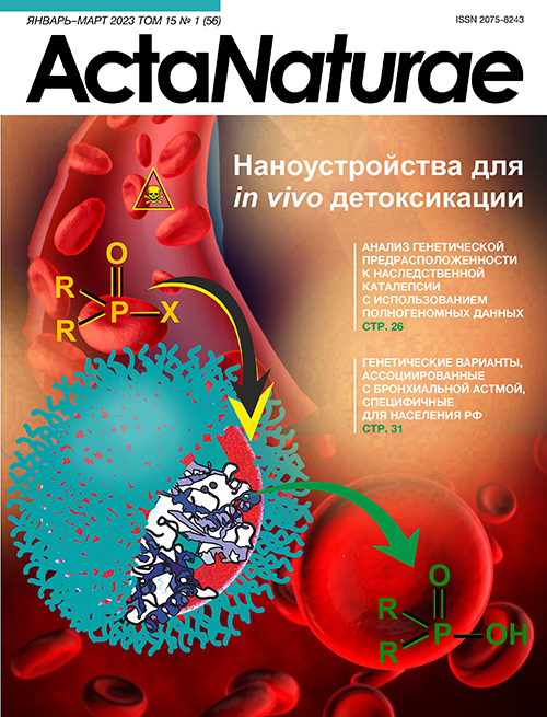
Наногубки. Наногубки представляют собой разлагающийся естественным образом трехмерный каркас, образованный в растворе небольшими молекулами, называемыми сшивающими агентами [54]. Впервые подход с наночастицами-наногубками, покрытыми природной клеточной мембраной и функционирующими посредством биомимикрии (рис. 2), был предложен Zhang L. [55]. «Наногубка действует как приманка для токсина in vivo и это новый способ удаления токсинов из кровотока», – сообщает Zhang L. «Вместо того, чтобы создавать специальные средства для лечения отдельных токсинов, мы разрабатываем платформу, которая может нейтрализовать токсины, продуцируемые широким спектром патогенов». Наногубка, созданная Zhang L. и его коллегами, представляет собой полимерное ядро из сополимера молочной кислоты с гликолевой кислотой (PLGA) с внешней оболочкой мембраны из эритроцитов, притягивающих токсины как приманка. В тестах на мышах профилактическое введение наногубок приводило к снижению уровня смертности до 11%, по сравнению со 100% уровнем смертности без лечения. Введение наногубок после инъекции токсина снижало смертность до 56%. Наногубки с изолированным токсином, предположительно, накапливались в печени, где в отсутствие каких-либо повреждений токсин безопасно метаболизировался и удалялся из организма [55, 56].

Рис. 2. Структура наногубки, представляющая собой полимерное ядро с мембранной оболочкой из эритроцитов. Адаптировано из [55]
Наногубки эффективно применялись для детоксикации бактериальных токсинов [57, 58]. Кроме того, такие наносистемы эффективно связывают и нейтрализуют низкомолекулярные соединения [59], аутоиммунные антитела [60], воспалительные цитокины [61], бактерии и вирусы [62, 63], нейротоксины (тетродотоксин, ботулинический токсин и сакситоксин) [64]. Наногубки, обеспечивающие нейтрализацию нейротоксинов, состоят из полимеров, покрытых мембраной нейронов, а именно клеток Neuro-2a; применение этой клеточной линии, полученной из нервного гребня мыши, повышало выживаемость мышей при лечении и профилактике при отсутствии острой токсичности [64]. Более эффективным был механизм двухмодальной детоксикации с наногубками, содержащими масляное ядро и покрытие эритроцитарной мембраной (Oil-NS) [65]. Полученная конструкция Oil-NS сочетает в себе специфическую связывающую способность биологических рецепторов, присутствующих на клеточной мембране, с неспецифической абсорбционной функцией масляного ядра, которые совместно повышают общую детоксикационную способность. Таким образом, нейтрализация токсинов осуществляется благодаря совместной работе мембраны и масляного ядра. Гибридная система наногубка-гель способна нейтрализовать токсины, а ее применение как в терапевтических, так и в профилактических целях приводит к значительному улучшению при поражении кожи токсинами [66]. Предметом дальнейшего изучения является стратегия биомиметической детоксикации, основанная на создании наночастиц, покрытых мембраной тромбоцитов, перспективных в качестве дополнительной терапии пациентов с инфекцией MRSA (метициллинрезистентный золотистый стафилококк) [67].
Эритролипосомы. Эритролипосомы (RM-PL) представляют собой биомиметическую платформу, сконструированную из искусственных липидных мембран и природных мембран эритроцитов. Такие системы успешно применяют для нейтрализации различных гемолитических порообразующих токсинов [68]. Токсины, поглощенные RM-PL, попадали в печень и селезенку, а затем подвергались эндоцитозу и перевариванию макрофагами. В результате утрачивалась первоначальная токсичность для органов-мишеней, что позволяло животным выжить.
Биомиметические гибридные системы. Микромоторы Янус – частицы магния и золота, покрытые мембранами эритроцитов (RBC-Mg), действующие как приманки и обладающие способностью поглощать и нейтрализовать биологические токсины в воде и биологических средах. Показана возможность применения наномоторов RBC-Mg для быстрой детоксикации α-токсина и метилпараоксона – моделей мембраноповреждающих токсинов и боевых отравляющих веществ соответственно [69, 70]. Гибридные биомембранные нанороботы с акустическим приводом и мембраной, состоящей из двух типов клеток (эритроциты и тромбоциты), эффективно связывались и c токсинами, и с патогенами в крови. Для одновременного элиминирования патогенных бактерий и токсинов были применены белки, находящиеся в гибридной мембране. Последние способствовали связыванию с патогенами и нейтрализации порообразующих токсинов [71, 72]. Опубликованы примеры [73] получения микророботов с наночастицами Fe3O4, покрывающими дрожжевые клетки, и создания цеолитного имидазолатного каркаса-67 (ZIF-67) для нейтрализации микотоксинов (рис. 3).

Рис. 3. Схема получения биомиметических гибридных систем для нейтрализации микотоксинов. Адаптировано из [73]
Нанодиализные системы для улучшения выведения токсинов
Липосомы. Использование липосомальных диализатов – это зарождающаяся область исследований. Липосомы, не загруженные лекарством, – «пустые» липосомы, использовали в качестве поглотителей экзогенных и эндогенных токсичных молекул, и часть из этих исследований достигла клинических испытаний. Вполне возможно, что в следующем десятилетии липосомы будут использоваться в качестве наноантидотов в клинике [74]. При введении «пустых» липосом in vitro происходит образование резервуара для связывания токсина. Липосомы обладают способностью связывать токсин за счет электростатических взаимодействий и гидрофобного эффекта в мембране или посредством захвата ионов в гидрофильном ядре. Внелипосомальные неионизированные молекулы проникают в липосомы и захватываются гидрофильным ядром с регулируемым значением рН. Например, слабоосновная лекарственная молекула, попадая в гидрофильное ядро липосомы с кислым значением pH, ионизируется и теряет способность диффундировать через липидный бислой (рис. 4).

Рис. 4. Схематическое изображение липосом, имеющих градиент рН между внутренней и внешней средой липосом (значения рН кислые/основные – внутри, нейтральный – снаружи). Адаптировано из [82]
Впервые гемодиализный метод, включающий липосомы и антиоксиданты, был представлен как уникальная стратегия удаления токсинов. In vitro наблюдали более заметное снижение содержания продуктов окисления и удаления тромбоцитов и билирубина по сравнению с обычным гемодиализом [75]. Эксперименты in vivo на крысах с уремией подтвердили, что добавление липосом в диализат в качестве дополнения к обычному гемодиализу может способствовать удалению связанных с белком уремических растворенных веществ. Разработанная наносистема обладает уникальными преимуществами в сравнении с альбумином и другими альтернативными методами с применением сорбентов [76]. Липосомы, модифицированные линолевой кислотой [77], и декорированные полиэтиленимином, продемонстрировали значительно более высокие скорости связывания и быстрый клиренс уремических токсинов, связанных с белками [78]. Доклиническая оценка трансмембранных липосом с градиентом рН для концентрирования аммиака подтвердила способность перитонеального диализа с липосомами снижать уровень аммиака в плазме у свиней с искусственно индуцированной гипераммониемией [79].
Перитонеальный диализ с липосомами (LSPD), а именно диализат, обогащенный рН-градиентными липосомами, т.е. имеющими градиент рН между внутренней и внешней средой липосом (кислая – внутри, нейтральная – снаружи), облегчал симптомы отравления на моделях животных [80, 81]. На крысах показано заметное повышение концентрации галоперидола, верапамила и амитриптилина в диализате при использовании LSPD по сравнению с перитонеальным диализатом без аугментации [80, 81]. LSPD применяли для удаления токсинов – препаратов, обладающих способностью сильно связываться с белками крови. Амитриптилин был выбран в качестве препарата, обладающего высокой способностью связываться с белками крови. Установлено, что диализаты, обогащенные липосомами, увеличивают экстракцию амитриптилина in vivo [82].
Липосомы, модифицированные полиэтиленгликолем с инкапсулированным фосфатсвязывающим цитратом железа (III), представляли собой ловушки для ионов фосфата в кровотоке во внутреннем липосомальном ядре и снижали концентрацию свободных ионов фосфата в растворе и в сыворотке [83] (табл. 1).
Таблица 1. Типы наноустройств для детоксикации, тип материала и библиотека ферментов / лекарственных веществ
Наноустройство | Материал | Нейтрализация | In vivo модель | Ссылка |
ЛЭ | Липоамин | Карго-альдегиды | - | [34] |
Интралипид | Пропранолол | Белые кролики | [36] | |
Интралипид | Кокаин | Клинические | [37] | |
Интралипид | Кокаин | Собака | [38] | |
Интралипид | Дилтиазем | Клинические | [39] | |
Интралипид | Бупренорфин, фентанил, буторфанол | - | [40] | |
Интралипид | Бупивакаин | Свиньи | [41] | |
Интралипид | Ивермектин | Pogona vitticeps | [42] | |
Интралипид | Ропивакаин | Свиньи | [44] | |
Интралипид | Севофлуран, изофлуран | Крысы | [45] | |
Интралипид | Амоксапин | Клинические | [46] | |
Интралипид | ФОС | Клинические | [48] | |
Нанокапсулы | Полисилоксан, октадецилтриметоксисилан, этилбутират, лецитин, Tween-80 | Хинолин | - | [52] |
Акриламид, N-(3-аминопропил)-метакриламид, N,N’-метиленбисакриламид, ферменты (алкооксидаза, каталаза,альдегид-дегидрогеназа) | Этанол | Мыши C57BL/6 | [53] | |
Наногубки | Мембрана эритроцитов, PLGA | Бактериальные токсины (α-гемолизин, листериолизин O, стрептолизин O) | - | [57] |
Мембрана эритроцитов, PLGA | Бактериальные токсины | Мыши CD-1 | [58] | |
Мембрана эритроцитов, PLGA | Дихлофос | Мыши CD-1 | [59] | |
Мембрана эритроцитов, PLGA | Аутоиммунные антитела | Мыши CD-1 | [60] | |
Мембрана нейтрофилов, PGLA | Воспалительные цитокины | Мыши ICR | [61] | |
Бактериальная мембрана, PLGA | Бактерии | Мыши C57BL/6 | [62] | |
Мембрана легочных эпителиальных клеток/мембрана макрофагов, PLGA | SARS-CoV-2 | Мыши C57BL/6NHsd | [63] | |
Мембрана клеток Neuro-2a, PLGA | Тетродотоксин | Мыши ICR | [64] | |
Мембрана эритроцитов, оливковое масло | ФОС (параоксон, диизопропил флуорофосфат, дихлофос) | Мыши ICR | [65] | |
Мембрана эритроцитов, PLGA, Pluronic F127 | Порообразующие токсины | Мыши ICR | [66] | |
Мембрана тромбоцитов, PLGA | S. aureus | Мыши CD-1 | [67] | |
Эритролипосома | Мембрана эритроцитов, холестерин,фосфатидилхолин, mPEG-DSPE | Порообразующие токсины | Мыши ICR | [68] |
Микромоторы Янус | Мембрана эритроцитов, Mg, Au, хитозан | α-токсин | - | [69] |
Мембрана эритроцитов, Au,лимонная кислота | Мелиттин | - | [70] | |
Гибридные биомембранные нанороботы | Мембрана эритроцитов, Au | Порообразующие токсины | - | [71] |
Мембрана эритроцитов и тромбоцитов, Au | Порообразующие токсины | - | [72] | |
Микророботы Янус | Мембрана дрожжевых клеток, Fe3O4,2-метилимидазол | Микотоксины | - | [73] |
Липосомы | Лецитин, холестерин, дезоксихолат натрия | Связанные с белкамиуремические токсины | Крысы Sprague Dawley | [76] |
Лецитин, холестерин, Tween-80,линолевая кислота | Связанные с белкамиуремические токсины | - | [77] | |
Лецитин, холестерин, линолевая кислота,полиэтиленимин, Tween-80 | Связанные с белкамиуремические токсины | - | [78] | |
LSPD | DPPC, холестерин, mPEG-DSPE,лимонная кислота | Аммиак | Минипиг Göttingen | [79] |
DPPC, холестерин, DSPE-mPEG | Аммиак | Крысы Sprague Dawley | [80] | |
DPPC, холестерин, DSPE-mPEG | Амитриптилин | Крысы Sprague Dawley | [81] | |
DOPG, холестерин | Амитриптилин | Крысы Sprague Dawley | [82] | |
Фосфатидилхолин, холестерин,DSPE-mPEG, цитрат железа | Ионы фосфата | - | [83] | |
DOPE-NHS, β-октилглюкозид, ферменты (алкогольоксидаза, каталаза) | Этанол | Крысы Sprague Dawley | [84] |
ФЕРМЕНТАТИВНЫЕ НАНОУСТРОЙСТВА ДЛЯ ДЕТОКСИКАЦИИ
Подробное описание наночастиц с инкапсулированными ферментами представлено в недавно опубликованном нами обзоре, где рассмотрены типы и материал наночастиц, результаты клинических исследований и т.д. [85]. Поэтому в этой части обзора сосредоточимся на системах – ферментативных наноустройствах для нейтрализации токсинов. Инкапсулирование ферментов в наноносители открывает возможность для создания наноустройств – нанореакторов, содержащих молекулы, осуществляющие аномальную диффузию и кинетические законы. Такие системы способны осуществлять одиночные и/или каскадные реакции, биосинтез, деградацию токсичных молекул [86]. Нанобиотехнология ферментных нанореакторов – это новая, активно развивающаяся область исследований. Например, недавно была исследована возможность перитонеального диализа с липосомами, содержащими ферменты (алкогольоксидазу и каталазу). Удаление этанола дополнительно ускорялось добавлением H2O2, который быстро разлагался до O2 с помощью каталазы. В модели интоксикации грызунов этанолом ферментные липосомы усиливали метаболизм этанола, о чем свидетельствовала повышенная продукция основного метаболита этанола – ацетальдегида [84].
Работы нашей группы сосредоточены, в частности, на проектировании и разработке инъекционных терапевтических ферментных нанореакторов для нейтрализации таких токсинов, как фосфорорганические соединения (ФОС) [87]. Известно, что ферменты, способные нейтрализовать ФОС, могут быть использованы в качестве стехиометрических, псевдокаталитических или каталитических «биоловушек» [88, 89]. Такие ферменты, как фосфотриэстеразы и холинэстеразы, могут быть активными компонентами терапевтических наноустройств. Инкапсулирование ферментов в наноносители предназначено, в первую очередь, для преодоления быстрого клиренса и иммунного ответа после инъекции гетерологичных терапевтических ферментов. Во-вторых, инкапсулирование фермента обеспечивает высокую концентрацию каталитического фермента в стабильных наноконтейнерах. Определение концентрации инкапсулированного фермента внутри наноносителей является важным этапом при конструировании эффективного нанореактора in vivo. В условиях инъекционного нанореактора токсикант, присутствующий в кровотоке, диффундирует через мембрану нанореактора, где далее в его герметичном отсеке проходит ферментативная реакция детоксикации [90]. Концентрация фермента (E) внутри наноносителя может быть как низкой, так и значительно превышать концентрацию токсиканта (Т). Реакция внутри нанореактора протекает в условиях либо первого ((E) << (T)), либо второго порядка, когда (E) ≈ (T). Возможны случаи, когда происходит лишь частичная инкапсуляция фермента, и на внешней поверхности нанореакторов образуется ферментная «корона», что может осложнить процесс и привести к нежелательному быстрому клиренсу и возможным неблагоприятным иммунным ответам на гетерологичные ферменты. Таким образом, проницаемость мембраны нанореактора для субстратов и продуктов реакции, возможные осмотические эффекты, эффекты вязкости и краудинга, образование ферментативной короны являются важными, еще не до конца решенными технологическими проблемам [90].
ЗАКЛЮЧЕНИЕ
В настоящее время наблюдается рост числа публикаций, посвященных созданию альтернативных, эффективных, многофункциональных, биомедицинских систем, таких, как роботизированные наноустройства для детоксикации. Обзор опубликованных данных показывает, что для доказательства выдвинутой концепции нанодетоксикации необходимы, прежде всего, междисциплинарный подход и объединение знаний в области создания и технологии наносистем, биохимии, биотехнологии, микро- и оптоэлектроники и т.д. Тем не менее, одним из возможных направлений в терапии острых отравлений является создание наномедицинских препаратов без наполнителей, состоящих из материалов и веществ, одобренных к клиническому применению. Кроме того, использование наноустройств открывает новые возможности для детоксикации при бактериальных и вирусных инфекциях.
Однако необходимо преодолеть еще долгий путь для создания высокочувствительных, легкоуправляемых и безопасных наноустройств и решить такие проблемы, как движение в узких и труднодоступных местах, например, капилляры кровеносных сосудов, выполнять сложные функции, быть гибкими и экономически эффективными.
Работа Т.Н. Пашировой, З.М. Шайхутдиновой и В.Ф. Миронова выполнена за счет государственного задания ФИЦ КазНЦ РАН.
Работа П. Массона (Ферментативные устройства для детоксикации) выполнена за счет средств субсидии, выделенной в рамках государственной поддержки Казанского (Приволжского) федерального университета в целях повышения его конкурентоспособности среди ведущих мировых научно-образовательных центров.
Дополнительные файлы

