


О биоразнообразии микробиома воздуха
- Авторы: Наумова Н.Б.1, Кабилов М.Р.1
-
Учреждения:
- Институт химической биологии и фундаментальной медицины СО РАН
- Выпуск: Том 14, № 4 (2022)
- Страницы: 50-56
- Раздел: Обзоры
- Дата подачи: 28.12.2021
- Дата принятия к публикации: 11.11.2022
- Дата публикации: 20.01.2023
- URL: https://actanaturae.ru/2075-8251/article/view/11671
- DOI: https://doi.org/10.32607/actanaturae.11671
- ID: 11671
Цитировать

Полный текст
ВВЕДЕНИЕ
Микроорганизмы повсеместно встречаются в окружающей среде и играют ключевую роль практически во всех экосистемах [1]. В связи с распространением многих патогенных микроорганизмов воздушно-капельным путем, в том числе и коронавируса SARS-CoV-2, вызвавшего текущую пандемию COVID-19, изучение, мониторинг и регулирование состава воздуха как снаружи, так и внутри помещений приобрели особую актуальность [2, 3]. К настоящему времени уже накоплено много информации о корреляции между загрязнением наружного воздуха и более тяжелым течением COVID-19: так, в Индии более низкий уровень смертности от заболевания наблюдали в городах с лучшим качеством воздуха [4]. Напомним, что термин «биоаэрозоль» охватывает широкий спектр органических частиц, содержащихся в атмосфере, источником которых являются разнообразные живые и мертвые организмы [5]. Как правило, наряду с частицами микробного, растительного или животного происхождения биоаэрозоли также содержат широкий спектр антигенных соединений, микробных токсинов и вирусов [6, 7]. Понимание процессов образования биоаэрозолей, закономерностей их распределения, распространения, перемещения, структуры и т.п., особенно в экстремальных условиях верхних слоев атмосферы, необходимо для широкого ряда фундаментальных и прикладных научных дисциплин [8], таких, как физика и химия, метеорология, гидрология атмосферы; изучение содержания аллергенных частиц и микроорганизмов, патогенных для человека, сельскохозяйственных животных и растений; а также аэробиология, биогеография и биоразнообразие, общая экология в целом. Основными направлениями изучения биоаэрозолей являются: а) оценка их источников и потоков; б) пространственное распределение и его изменение во времени; в) старение биологических частиц; г) метаболическая активность; д) урбанизация аллергий; е) транспорт патогенов и ж) влияние на климат [8].
Цель этого обзора – краткое описание микробиоты биоаэрозолей, с акцентом на составе и структуре микробиома. Воздух является исключительно динамичной и, как следствие этого, очень проблематичной средой для отбора и анализа образцов биоаэрозолей, установления источников аэрозолизации и путей переноса, поэтому методологические аспекты сбора образцов, вне всякого сомнения, имеют огромное значение для интерпретации и сравнения данных. Большое значение имеют и методы анализа микробиома. Тем не менее, так как эти два направления являются обширными, мы коснемся их в этом обзоре лишь кратко.
ОСНОВНЫЕ СВОЙСТВА БИОАЭРОЗОЛЕЙ
Биоаэрозоль является важной частью атмосферного аэрозоля. Расчеты показывают, что среди представленных в воздухе частиц биоаэрозоль занимает по объему 10–28% [9], а по массе 16–80% [1].
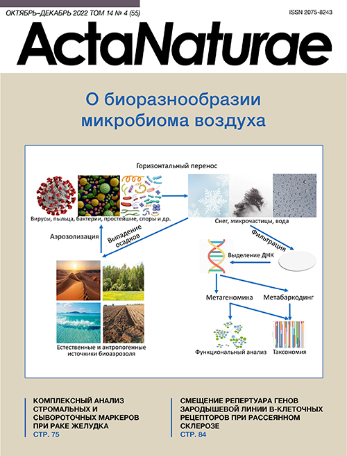
Распространение микроорганизмов по воздуху происходит повсеместно, и у некоторых из них является важной частью жизненного цикла [10]. В образование биоаэрозолей вносят вклад различные природные источники (рис. 1) – почва, лес, пустыни, океаны и моря и т.д. [11], а также антропогенные (сельское хозяйство, пищевая промышленность, свалки и т.д.) [6, 11, 12].

Рис. 1. Схематическое изображение образования, распространения и анализа биоаэрозолей
После попадания в атмосферу, т.е. аэрозолизации, микроорганизмам в гораздо большей степени по сравнению с их индигенными местообитаниями – источниками аэрозоля – приходится испытывать стресс иссушения, УФ-облучения, низких температур и низкого содержания источников углерода и энергии, и многие могут не выдержать [13].
Размеры биоаэрозольных частиц варьируют от 3 нм [14] до 100 мкм в зависимости от источника происхождения: диаметр пыльцы 17–58 мкм, спор грибов 1–30 мкм, клеток бактерий обычно 0.25–8 мкм [15], а вирусов меньше 0.3 мкм. Причем совершенно не обязательно, что биологический материал представлен в виде отдельных частиц: большинство бактерий связано с частицами с диаметром более 2 мкм [16, 17], 2–3 мкм [18], 3–4 мкм [19, 20]. В некоторых случаях выявлено бимодальное распределение бактерий по размерам частиц биоаэрозоля с одним пиком 1–2 мкм и вторым в области 4–7 мкм [21]. Бактерии также могут встречаться в виде агломератов клеток или быть связаны с частицами растений, животных или почвы, а также с пыльцой или спорами. Переносимые в атмосфере клетки бактерий и споры грибов могут достигать концентраций ~103÷104 и ~105 в 1 м3 [17, 21] и встречаться на высоте вплоть до 40 км над уровнем моря, т.е. до стратосферы [22]. Концентрация бактериальных частиц, способных образовывать колонии на лабораторных питательных средах, в приземном слое тропосферы в городских условиях на юге Польши варьировала от 65 до 355 КОЕ/м3 [19] и в пределах 300–1350 КОЕ/м3 в городской и сельской местности в Таиланде [18]; при этом в последнем случае число КОЕ быстро падало с высотой – в 2 раза при переходе от 1–3 до 7 м над поверхностью суши. В приземных и более высоких слоях (несколько тысяч метров) тропосферы над югом Западной Сибири лабораторное культивирование выявило значительное преобладание спорообразующих бактерий Bacilli/Firmicutes [23, 24], в то время как над севером региона преобладали неспорообразующие бактерии [25].
Распределение биоаэрозолей в воздухе зависит от времени года [19, 26]. Так, в воздухе приморского района Китая концентрация клеток бактерий, определенная путем микроскопирования, зимой была выше, чем летом [21]. Нагрузка биоаэрозолей патогенной микробиотой может сильно меняться в зависимости от времени года: так в Южной Азии было выявлено существенное повышение содержания патогенов в постмуссонный период и зимние месяцы. Выявлено и значительное суточное варьирование состава биоаэрозолей [26].
Статистически наиболее значимыми метеорологическими факторами, определяющими жизнеспособность переносимых воздухом бактерий, являются температура и ультрафиолетовое облучение [19, 21]. Биотическая нагрузка аэрозолей и их поведение в окружающей среде в значительной степени зависят от загрязнения воздуха (дымка, туман, пыль, разные макрочастицы), в том числе транспортом и сжиганием биомассы [26]. При этом доля жизнеспособных бактерий в общем пуле может варьировать в зависимости от степени загрязнения [15]. Состав биоаэрозолей может меняться в зависимости от специфических случайных метеорологических условий: пыльные бури, например, приводят к сильному увеличению концентрации микроорганизмов в биоаэрозоле [16], при этом разные компоненты биоаэрозолей по-разному изменяются в зависимости от метеоусловий.
Накопленная к настоящему моменту информация свидетельствует о важной роли биоаэрозолей [6, 11, 27, 28] в физических и химических процессах, протекающих в атмосфере [1, 29]. Показано, что биоаэрозоли могут присоединяться к окружающим частицам и таким образом оказывать влияние на атмосферные процессы, выступая ядрами конденсации в облаках и инициируя выпадение осадков [10, 30, 31]. Так, установлено, что в 33% случаев именно биологические частицы служили ядрами при образовании снега и облаков [32].
Наряду с воздействиями на погодные явления, биоаэрозоли влияют и на здоровье людей [33], так как в их состав могут входить патогенные/условно патогенные бактерии, грибы, вирусы, высокомолекулярные аллергены, бактериальные эндотоксины, микотоксины, пептидогликаны, бета-(1–3)-гликаны, пыльца и растительные волокна [6]. В первую очередь неблагоприятное воздействие биоаэрозоля на здоровье человека проявляется в респираторных симптомах. Например, показана высокая корреляция между повышением концентрации пыльцы на открытом воздухе весной и летом и обострением астмы у детей [34]. Установлена связь между содержанием грибных спор в воздухе и частотой обращения пациентов с астматическими симптомами [35]. Эндотоксин бактериальных биоаэрозолей признан важным этиологическим фактором профессиональных заболеваний легких, включая астму (неаллергическую) [6]. Изоляты Escherichia coli, которые обычно используют в качестве индикаторов качества воды, обнаружены также и в атмосферной пыли [36].
СБОР ОБРАЗЦОВ АЭРОЗОЛЯ
Сбор образцов аэрозоля основан на различных физических подходах отделения частиц от воздушного потока [37]. Но общий смысл заключается в прокачивании насосом воздуха через фильтр или жидкую среду, улавливающую аэрозольные частицы [38]. Недавно стали появляться методы, позволяющие в процессе сбора образцов разделять частицы по размеру [39]. Это особенно актуально для аэровирусологии – в последнее десятилетие активно разрабатываются методы сбора образцов аэрозоля внутри помещений для мониторинга влияния дыхания людей. В целом инструментальные варианты сбора аэрозоля пока не стандартизированы и сильно варьируют, однако общий принцип их работы остается неизменным.
МЕТАГЕНОМНОЕ СЕКВЕНИРОВАНИЕ
В настоящий момент изучение таксономического разнообразия микробиоты биоаэрозоля основано на подходах, использующих высокопроизводительное секвенирование. Из всей совокупности уловленных фильтром или жидкой средой микроорганизмов извлекают тотальную ДНК, которую далее используют в метагеномном анализе. С развитием таких методов стала возможной идентификация некультивируемых микроорганизмов, составляющих основу воздушного биома [40]. Полученные к настоящему времени результаты метагеномных исследований показали, что доминантные виды идентифицированных таким образом микроорганизмов отличаются от доминантов, выявленных при стандартном культивировании [41], поскольку более 99% микроорганизмов, обнаруженных в воздухе, не растут в лабораторных условиях [26]. Появилось и стало широко использоваться понятие «микробиом», которое в одноименном журнале (Microbiome) определяют так: «Этот термин относится ко всему местообитанию, включая все микроорганизмы (бактерии, археи, низшие и высшие эукариоты, и вирусы), их геномы (т.е. гены) и условия окружающей среды» [42]. Однако опубликованные работы, в заголовках или ключевых словах которых есть слово «микробиом», не следуют этому определению, так как подавляющее большинство исследований сфокусированы только на одной группе (вирусы, бактерии, грибы или растения), в лучшем случае на комбинации двух. Не вдаваясь в причины такого положения дел, здесь мы просто ограничимся констатацией этого и подчеркнем, что далее под «микробиомом» мы будем иметь в виду – вслед за авторами цитируемых работ – бактериальную или грибную составляющие микробиома, либо их комбинацию.
Итак, можно найти огромное количество публикаций, связанных с изучением микробиомов всевозможных природных объектов, например, горячих источников, озер, морей, почвы, эндогенной микробиоты организмов и т.д. [43–46], однако метагеномный анализ биоаэрозолей представлен катастрофически меньшим числом работ [47–52].
Условно метагеномное секвенирование можно разделить на два глобальных направления: полногеномное секвенирование (метагеномика) и таргетное (метабаркодинг). В первом случае идет прочтение всей ДНК, выделенной из образца, что позволяет, с одной стороны, говорить о таксономическом разнообразии, а с другой – предоставляет возможность анализа функциональных свойств. Однако стоимость метагеномного подхода заведомо выше [48, 53], чем метабаркодинга, который основан на анализе высококонсервативных маркерных генов, таких, как 16S (бактерии, археи), ITS (грибы, растения), rbcL (растения), 18S (различные эукариоты) и др. [54, 55]. При этом эффективность таксономической идентификации напрямую зависит от количества верифицированных последовательностей в используемых специализированных базах данных. Наиболее полными в настоящий момент являются базы прокариот (16S) и грибов (ITS).
Исключительно низкое содержание микроорганизмов в воздухе, наряду с сильным варьированием состава микробных ансамблей, является серьезной проблемой для анализа биоразнообразия, функционального спектра и метаболической активности микробиоты биоаэрозолей [56]. Подчеркнем, что исследования такого типа являются фундаментальной основой для выявления аспектов взаимодействия человека с природой, в частности, касающихся способов передачи заболеваний и потенциального воздействия на здоровье человека [57]. Тем не менее, опубликованы только единичные результаты метагеномного анализа биоаэрозолей в России [58].
Бактериобиом биоаэрозолей
В приповерхностных слоях атмосферы бактерии составляют существенную часть биоаэрозолей: так, в горах Колорадо (США) бактерии в среднем составляли 22% от аэрозольных частиц размером более 0.5 мкм [47].
Бактерии аэрозолей могут значительно влиять на химию атмосферы, оказывая воздействие на здоровье человека [15]. Так, высокий уровень загрязнения воздуха может очень сильно изменять структуру его бактериобиома [59]. В туманные дни в Пекине выявлено повышение содержания патогенных бактерий Halomonas и Shewanella [60], особенно осенью и ранней зимой.
Большое биоразнообразие бактериобиома биоаэрозолей установлено путем метагеномного секвенирования [61]. Например, в приземных слоях тропосферы в условиях города идентифицировали бактерии 38 таксономических типов [41]. В большинстве исследований установлено, что Proteobacteria, Firmicutes и Actinobacteria являются основными доминантами бактериобиома нижних [41, 62, 63] и более высоких слоев тропосферы [50, 64, 65], при этом в нижних слоях в городских условиях Firmicutes могут вносить заметный (20–30%) вклад, а такие типы, как Cyanobacteria, Bacteroidetes, Chloroflexi, Acidobacteria и Deinococcus-Thermus, выявляют как минорные (1–5% относительного содержания нуклеотидных последовательностей) доминанты. Однако другими исследованиями показано высокое содержание представителей Bacteroidetes в биоаэрозоле над Японией после пылевых бурь в Азии [17, 66], а также в воздухе над восточной Австралией [67]. Весьма специфичным был состав бактериобиома в высоких слоях тропосферы над полуостровом Ното в Японии, где (правда, методом флуоресцентной гибридизации in situ) показано, что 80% всех эубактерий на минеральных частицах аэрозоля были представлены Bacillus subtilis, относящейся к фирмикутам [68].
Различные метеорологические события оказывают значительное влияние на состав и структуру бактериобиома биоаэрозолей. Например, перенос воздушными потоками пылевых частиц, аэрозолизированных пыльными бурями, на дальние расстояния над морями и континентами является важным механизмом попадания в местные экосистемы различных микроорганизмов [69]. Так, бури в Сахаре приводят к попаданию пылевых частиц в атмосферу, затем вместе с движением воздушных масс эти частицы переносятся в Европу, приводя, в частности, к их накоплению в снеговом покрове Альп на высоте более 3000 м над уровнем моря [70]. Биоиндикаторами пылевых частиц из Алжира являлись представители Gemmatimonadetes и Deinococcus-Thermus [70], известные своей встречаемостью в сухих олиготрофных местообитаниях с относительно высоким уровнем солнечной радиации, что и позволяет им выживать в процессе такой транспортировки, сохраняя при этом метаболическую активность. Хотя и в очень низких количествах, но патогенные бактерии могут тоже переноситься с пылевыми частицами на очень большие расстояния [70]. При этом поверхность тела человека является более вероятным (по сравнению с другими биотопами) источником патогенных бактерий в воздухе [71]. Установлена четкая зависимость структуры и состава бактериобиома на высоте 10 м от поверхности суши (остров и полуостров в Восточной Азии) от пылевых бурь в Центральной Азии [69]. При этом пылевые частицы служат центрами нуклеации льда [72]. Выпадение осадков является не менее важным механизмом привноса микроорганизмов из верхних в нижние слои тропосферы и на поверхность территорий [73]. Это исследование показало, что состав бактериобиома в осадках а) соответствовал источникам биоаэрозолей на пути переноса и б) имел выраженную сезонную динамику со снижением относительного обилия превалирующих Proteobacteria от лета к зиме.
Примечательно, что, в отличие от микобиома, состав которого внутри помещений определялся его составом снаружи и не зависел от активности людей внутри, биоразнообразие бактериобиома внутри помещений зависело как от бактериобиома снаружи [74], так и от активности людей внутри [41]. Однако загрязнение наружного воздуха может не влиять на биоразнообразие ансамблей бактерий и архей в биоаэрозоле внутри помещения, как показало исследование в Пекине [74]. Это свидетельствует о различных механизмах формирования и динамики разных составляющих микробиома, что следует иметь в виду при планировании соответствующих наблюдений.
Заметный вклад в суммарную нагрузку воздушных частиц могут вносить Cyanobacteria, вызывающие различные проблемы со здоровьем после вдыхания [75]. Недавно пикоцианобактерии были обнаружены в приповерхностных слоях атмосферы над землей или водоемами в Гренландии и Антарктике [76], где аэрозолизация почвы и воды является ведущим механизмом образования аэрозолей; их распространение ветром считается основным источником поступления Cyanobacteria в воздух.
Проведенный метаанализ результатов 42 исследований, в совокупности охватывающих более трех тысяч образцов биоаэрозоля, выявил повышенное разнообразие бактерий и относительное обилие патогенов в образцах, так или иначе ассоциированных с антропогенной активностью в местах сбора [71].
Микобиом биоаэрозолей
Микобиом аэрозолей сильно варьирует, но на уровне типа, как правило, основными компонентами микобиома как в приземных, так и в более высоких слоях тропосферы являются Basidiomycota и Ascomycota, меняющиеся местами в плане основного доминирования. Так, представители типа Ascomycota значительно (более двух третей) доминировали в приповерхностном слое воздуха в горах Колорадо на высоте более 3000 м над уровнем моря [77], а также и в приповерхностных воздушных слоях Кувейта на существенно более низкой высоте над уровнем моря [78]. Другие исследователи, однако, выявили доминирование типа Basidiomycota (до 60% и выше) [41, 63, 79], в то время как Ascomycota составляли около трети последовательностей грибов. Интересно, что присутствие устойчивых к атмосферным стрессам представителей Ascomycota (Cladosporium и Alternaria) увеличивалось с повышением высоты (500–800 м по сравнению с 5–10 м) над пустынями Гоби и Таклимакан [80], которые являются основными поставщиками пылевых частиц в атмосферу Азии. В приповерхностном воздухе над горой высотой 3043 м над уровнем моря в Австрии основными были представители классов Basidiomycota (Agaricomycetes), за которыми следовали представители таких классов аскомицетов, как Dothideomycetes, Saccharomycetes, Sordariomycetes, Leotiomycetes и Eurotiomycetes [64]. Аскомицеты – представители семейства Davidiellaceae – составляли 25% микобиома в направлении с северо-востока Китая в Японию [81]. Однако в одном из последних исследований биоразнообразия грибов в аэрозолях над Антарктикой представители этого семейства не были обнаружены среди доминантов микобиома [82]. Гриб Alternaria, относящийся к Pleosporaceae /Pleosporales / Dothideomycetes / Ascomycota, часто выявляют среди основных доминантов приземных слоев как в городских (Нандзин, Пекин, Сеул), так и в естественных (пустыня в Кувейте) условиях [41, 78, 83]. Хорошо известные как основные компоненты аэрозольной микобиоты культивируемые роды грибов Alternaria, Aspergillus, Penicillium, Cladosporium и др. [84] при метагеномном подходе могут составлять не более 12% от общего числа маркерных нуклеотидных последовательностей [41]. Однако следует иметь в виду, что относительное содержание Alternaria в воздухе может сильно варьировать (от 10 до 40%) в зависимости от года как над сельской, так и над городской местностью [85]. В этой же работе установлена зависимость состава микобиома приповерхностных аэрозолей от типа и состояния растительности (влажности листьев). Некоторые статьи описывают довольно неожиданный (в смысле – сильно отличающийся от данных других исследований) состав микобиома. Так, показано, что последовательности рода Candida (Saccharomycetales/ Saccharomycetes/ Ascomycota) составляли 54% микобиома нижнего слоя тропосферы [81]. Что касается приземного слоя, то в той же работе [81] установлено, что микобиом состоял исключительно из аспергилл (Aspergillus / Aspergillaceae / Eurotiales / Eurotiomycetes /Ascomycota). Очевидно, что состав биоаэрозолей внутри помещений существенно зависит от состава воздуха приземных слоев атмосферы снаружи и это особенно касается микобиома, состав которого, как показано в проведенном в Корее исследовании воздуха внутри помещений детских садов, определялся составом биоаэрозоля вне помещений и практически не зависел от активности людей [41]. Разнообразие микобиома внутри помещений может зависеть от загрязнения воздуха снаружи, как показано при исследовании в Пекине [74]. Как и в случае бактериобиома, состав микобиома может меняться в зависимости от конкретных метеорологических событий: так, после дождя над засушливой территорией Средиземноморья существенно повышалось содержание грибов класса Agaricomycetes/ Basidiomycota [86], которые после дождей выпускают огромное количество спор в воздух.
ЗАКЛЮЧЕНИЕ
Таким образом, микробиом биоаэрозоля представляет собой в высшей степени динамичную систему. Изменение состава и структуры микробиома зависит от огромного числа разных факторов, многие из которых опосредуют, маскируют, влияют на действия друг друга, так или иначе мешая установить четкие пространственно-временные закономерности. Перенос микроорганизмов на большие расстояния воздушными течениями в верхних слоях тропосферы в конечном итоге оказывает серьезное влияние на состав низких слоев, с которыми непосредственно контактирует человек. Это может иметь большое значение с точки зрения способов распространения некоторых заболеваний и потенциального воздействия на здоровье человека, особенно в условиях роста народонаселения планеты и загрязнения окружающей среды. Поэтому настоятельную необходимость укрепления позиций России в плане научного изучения и мониторинга воздушного пространства, в частности, микробиологической составляющей биоаэрозолей, нельзя переоценить.
Исследование выполнено при финансовой поддержке РФФИ в рамках научного проекта № 19-05-50032.
Дополнительные файлы

