


Искусственные хромосомы человека и способы их доставки в клетки-мишени
- Авторы: Пономарцев С.В.1, Синенко С.А.1, Томилин А.Н.1,2
-
Учреждения:
- Институт цитологии РАН
- Институт трансляционной биомедицины, Санкт-Петербургский государственный университет
- Выпуск: Том 14, № 3 (2022)
- Страницы: 35-45
- Раздел: Обзоры
- Дата подачи: 27.12.2021
- Дата принятия к публикации: 19.07.2022
- Дата публикации: 29.10.2022
- URL: https://actanaturae.ru/2075-8251/article/view/11670
- DOI: https://doi.org/10.32607/actanaturae.11670
- ID: 11670
Цитировать

Полный текст
СПИСОК СОКРАЩЕНИЙ ИХЧ – искусственная хромосома человека; ИХБ – искусственная хромосома бактерий; ИХД – искусственная хромосома дрожжей; MMCT – перенос хромосом, опосредованный микроклетками (Microcell-Mediated Chromosome Transfer); TAR – рекомбинация, ассоциированная с трансформацией; CENP-B – центромерный белок B; ЭСК – эмбриональные стволовые клетки; ИПСК – индуцированные плюрипотентные стволовые клетки; CHO – клетки яичника китайского хомячка; tetO – тетрациклиновый оператор; HSV-1 – вирус простого герпеса типа 1; HVJ-E – японский гемагглютинирующий вирус E; MLV – ретровирус лейкоза мышей; TACF – теломер-ассоциированная фрагментация хромосом; FSCT – перенос хромосом с помощью проточной сортировки; MWCF – слияние целых микроядерных клеток; iMCT – трансфекция изолированных метафазных хромосом; ПЭГ – полиэтиленгликоль; HPRT – гипоксантин-гуанин-фосфорибозилтрансфераза; FACS – проточная цитометрия (fluorescence-activated cell sorting); GFP – зеленый флуоресцентный белок.
ВВЕДЕНИЕ
Искусственные хромосомы человека (ИХЧ, англ. human artificial chromosomes, HAC) были получены, в первую очередь, в качестве экспрессионных векторных систем для переноса трансгенов в эукариотические клетки. На сегодняшний день разработано множество векторных систем, различающихся по основным характеристикам: (1) способности встраиваться в хромосомы хозяйских клеток или оставаться в эписомной форме, (2) генетической емкости, определяющей максимальный размер трансгена, и (3) способу доставки. Интегрирующие векторы встраиваются в ДНК клеток-хозяев, в результате чего они наследуются дочерними клетками. К недостаткам таких векторных систем относятся случайный характер их встраивания в геном, ассоциированный с риском инсерционного мутагенеза, а также эпигенетическим подавлением экспрессии трансгенов. Интегрирующими векторами являются линеаризованные плазмиды, а также векторные системы на основе ретровирусов [1–3] и транспозонов, таких, как piggy-Bac, Sleeping Beaty, Tol2 [4–6].
Неинтегрирующие векторы находятся в хозяйских клетках в эписомном состоянии. В процессе клеточного деления происходит неравномерное распределение таких векторов между дочерними клетками и их постепенная утрата. Подобные системы удобны для временной трансфекции клеток, однако они не подходят для длительной экспрессии трансгенов. Примерами таких векторных систем являются кольцевые плазмиды и векторы на основе аденовирусов, альфавирусов, герпесвирусов, бакуловирусов, поксвирусов и бактериофагов [1, 2, 7]. Важным параметром векторных систем является их емкость, определяемая максимальным размером встраиваемого трансгена. С помощью плазмид можно переносить трансгены длиной до 20 т.п.н. Использование векторов на основе транспозонов позволяет осуществлять доставку трансгенной ДНК размером до 9 т.п.н., в то время как векторные системы на основе вирусной ДНК могут вмещать трансгены до 150 т.п.н. [1, 2]. Существуют различные способы доставки экспрессионных векторных систем в клетки-мишени. Плазмиды и векторы на основе ДНК-транспозонов переносят с помощью кальций-фосфатной трансфекции, электропорации, липофекции, сонопорации, микроинъекции, магнитофекции, а также с помощью так называемой «генной пушки». Доставка векторов на основе вирусной ДНК, называемая трансдукцией, осуществляется посредством характерного для данного вируса механизма заражения клетки хозяина.
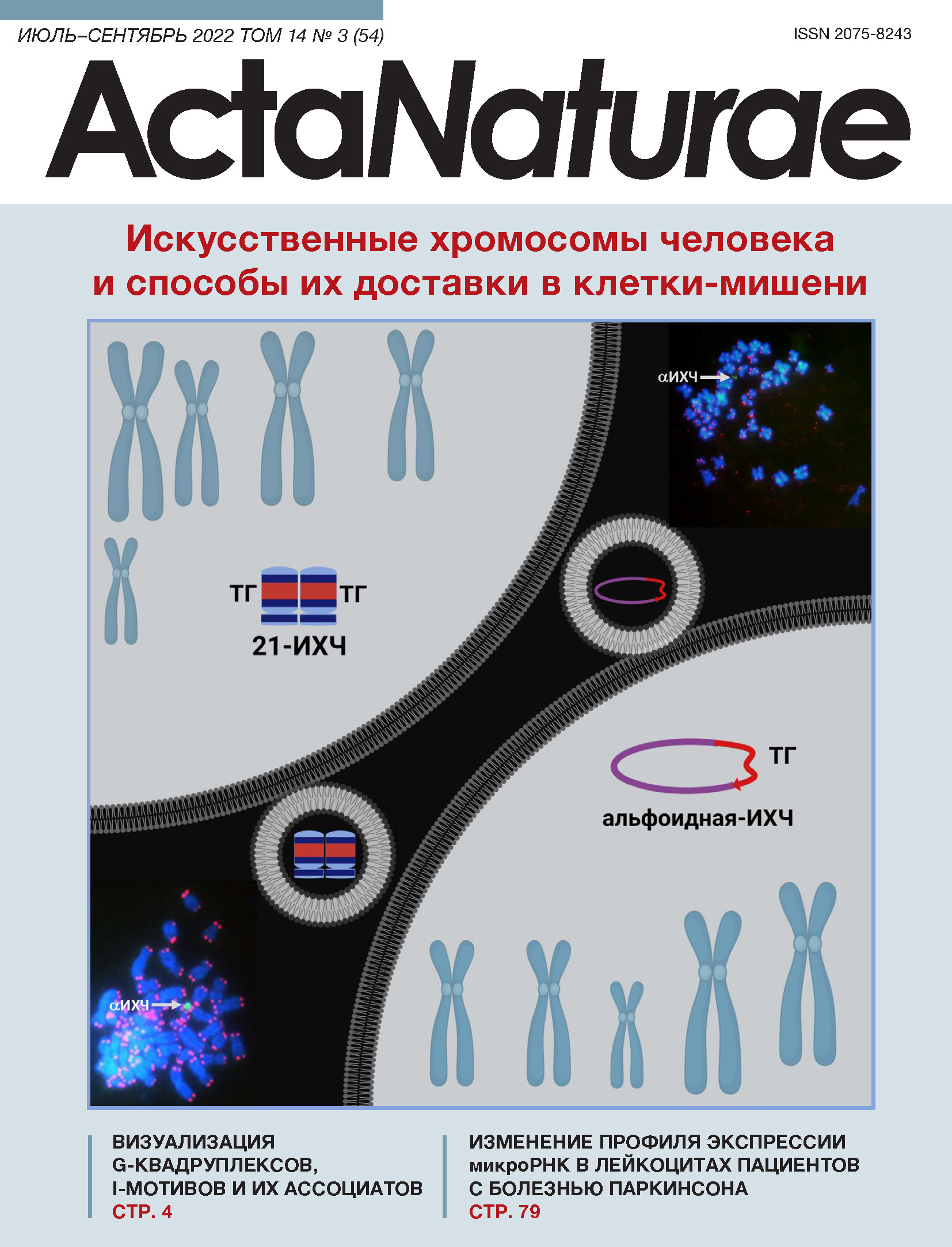
Искусственные хромосомы человека – это векторные конструкции, обладающие основными характеристиками хромосом: (1) способностью автономного существования в клетке как дополнительной хромосомы и (2) способностью реплицироваться и передаваться обеим дочерним клеткам в процессе клеточного деления. Таким образом, использование ИХЧ позволяет избежать рисков инсерционного мутагенеза и при этом обеспечивает устойчивую экспрессию трансгенов. Уникальной особенностью ИХЧ является их сверхвысокая емкость, позволяющая осуществлять перенос трансгенов длиной до нескольких миллионов пар нуклеотидов, в том числе целых генных локусов с цис-регуляторными участками, позволяя точно воспроизводить экспрессию эндогенных локусов. Хотя на сегодняшний день получено множество ИХЧ различной структуры, эти векторные системы продолжают интенсивно совершенствоваться и модифицироваться [8–13]. ИХЧ получают с помощью двух подходов. Первый – это так называемый подход «сверху-вниз» (англ. top-down), который позволяет получать ИХЧ из нативных хромосом путем их максимальной редукции, не затрагивающей только компоненты, необходимые для стабильной репликации в клетке, а именно, центромерный и теломерные участки [14–16]. Второй подход – синтетический, «снизу-вверх» (англ. bottom-up) – позволяет получать линейные или кольцевые ИХЧ посредством синтеза и сборки больших участков прицентромерной альфа-сателлитной ДНК in vitro [13, 17–19]. Необходимо отметить, что, несмотря на очевидные преимущества ИХЧ перед другими векторными системами, существует ряд технических ограничений для их активного использования как в научных исследованиях, так и в биомедицинских приложениях. Одно из главных ограничений – неэффективность и трудоемкость методов переноса ИХЧ в клетки-мишени. В нашем обзоре описаны разные типы ИХЧ, способы их доставки в клетки, а также перспективы внедрения этих эписомных векторов сверхвысокой емкости в практическую медицину.
ОСНОВНЫЕ ТИПЫ ИХЧ И СПОСОБЫ ИХ ПОЛУЧЕНИЯ
ИХЧ, получаемые путем редукции нативных хромосом человека
Укорочение хромосом эукариотических клеток стало возможным благодаря методу теломер-ассоциированной фрагментации хромосом (TACF, англ. Telomere-Associated Chromosome Fragmentation) [20]. На сегодняшний день такие ИХЧ являются наиболее охарактеризованными и усовершенствованными в контексте их использования в качестве стабильных экспрессионных векторных систем. Подход top-down позволяет удалять плечи хромосом, заменяя их новыми теломерсодержащими участками, которые встраивают в выбранные локусы с помощью гомологичной рекомбинации. В полученных таким образом ИХЧ могут оставаться единичные криптические гены и некодирующие последовательности, однако всегда присутствуют элементы, необходимые для их стабильного поддержания в клеточном ядре (теломеры) и равномерного распределения между дочерними клетками в процессе деления (центромеры). Чтобы осуществлять сайт-специфическое встраивание трансгенов в такие конструкции, в них предварительно вводят соответствующие последовательности, например, loxP-сайты, которые опосредуют интеграцию трансгенов посредством Cre-зависимой рекомбинации. Также, в ИХЧ часто помещают селективные маркеры, которые позволяют проводить позитивный отбор клеток, содержащих ИХЧ. С помощью метода TACF получены искусственные хромосомы на основе хромосом 14 [21] и 21 человека [16, 22, 23], а также хромосомы 11 мыши [24] – в последнем случае такие хромосомы называют искусственными хромосомами мыши. Наиболее технически продвинутой на сегодняшний день является ИХЧ на основе хромосомы 21 человека, или 21-ИХЧ [16], которую получали в несколько этапов (рис. 1). Нативную хромосому 21 человека сначала переносили в клеточную линию DT-40 курицы, удобную для осуществления гомологичной рекомбинации ДНК [25]. Далее с помощью метода TACF из перенесенной хромосомы удаляли p-плечо, для чего с помощью гомологичной рекомбинации в участок, находящийся вблизи центромеры, встраивали теломерную последовательность. Вместе с теломерной последовательностью встраивали также селективный маркер, который позволяет отобрать клетки, в которых произошла рекомбинация. Аналогичным образом удаляли и q-плечо (рис. 1). Помимо теломерного участка, в 21-ИХЧ был внесен также сайт loxP, фрагмент гена гипоксантин-гуанин-фосфорибозилтрансферазы (HPRT), а также другие элементы. С помощью секвенирования показано, что, помимо центромерного участка и встроенных элементов, полученная 21-ИХЧ содержит незначительное количество остаточного генетически инертного материала [26]. Полученную ИХЧ переносили из клеток линии DT-40 в клетки линии СНО яичника китайского хомячка, в которых затем проводили финальный этап сборки ИХЧ, заключающийся в «загрузке» требуемого трансгена с помощью сайт-специфической рекомбинации, а также поддержание и наработку этой ИХЧ. Затем ИХЧ переносили в клетки-мишени с помощью описанного ниже метода ММСТ (англ. Microcell-Mediated Chromosome Transfer).

Рис. 1. Схема сборки 21-ИХЧ с помощью метода теломер-ассоциированной фрагментации хромосом. Хромосому 21 из клеток человека переносили в клетки линии DT-40. Затем с помощью гомологичной рекомбинации вблизи центромеры в нее встраивали теломерные последовательности (отмечены синим), что приводило к фрагментации хромосомы в районе вставки. Таким образом, в результате последовательного удаления плеч хромосомы с их заменой на теломерные участки получали 21-ИХЧ (21HAC)
Создано несколько модификаций 21-ИХЧ, различия между которыми заключались в использовании различных селективных маркеров: гена зеленого флуоресцентного белка (GFP), гена тимидинкиназы (tk) вируса простого герпеса, а также генов устойчивости к неомицину, гигромицину и бластицидину [26]. Получена также 21-ИХЧ, содержащая мультиинтегразный локус, включающий сайты loxP, FRT, ФC31attP, R4attP, TP901-1attP и Bxb1attP [23]. Такую ИХЧ использовали в различных исследовательских и генотерапевтических моделях [8]. На основе этой ИХЧ созданы экспрессионные векторы для коррекции миодистрофии Дюшенна [22, 27, 28] и гемофилии А [29], с ее помощью проведено также репрограммирование эмбриональных фибробластов мыши [30].
ИХЧ на основе сателлитов
Еще один тип ИХЧ, получаемый по принципу top-down, образуется путем встраивания трансгена в кластер генов рибосомной ДНК на коротких плечах акроцентрических хромосом [31, 32] (рис. 2). При таком встраивании возможны ошибки репликации, в результате чего образуются длинные инвертированные повторы [33]. Наряду с этим происходит удвоение центромеры, хромосома «ломается», и фрагмент короткого плеча формирует отдельную хромосому, которая в дальнейшем ведет себя как самостоятельная репликативная единица [34, 35] и обеспечивает устойчивую экспрессию внедренного в нее трансгена [36]. Полученные таким способом ИХЧ, названные SATAC (от англ. Satellite-DNA-based Artificial Chromosome), выделяют из клеток-доноров с помощью проточной цитофлуориметрии, переносят в клетки-мишени с помощью дендримеров и катионных частиц [37], а также посредством микроинъекций [38, 39]. Эмбриональные стволовые клетки (ЭСК) мыши, в которые переносили SATAC, показывали способность участвовать в нормальном эмбриональном развитии [38, 40].

Рис. 2. Схема получения ИХЧ типа SATAC. В перицентромерную область акроцентрической хромосомы с помощью гомологичной рекомбинации вносят экзогенную ДНК, содержащую сайты для сайт-специфической рекомбинации, селективный маркер и другие последовательности. Вследствие такого встраивания происходит амплификация участков перицентромерной, рибосомной и экзогенной ДНК. После удвоения центромеры происходит фрагментация хромосомы, в результате чего формируется ИХЧ. Условные обозначения: Т – теломера, ПЛ – длинное плечо акроцентрической хромосомы, П – перицентромерный участок, Ц – центромера, Р – рибосомная ДНК, Э – экзогенная ДНК
Альфоидные ИХЧ
Принципиально иной способ создания ИХЧ основан на синтезе протяженных нуклеотидных последовательностей, обладающих основными функциями хромосом. Основную сложность в случае подхода bottom-up представляет конструирование функциональной искусственной центромерной последовательности. Как известно, в хромосомах человека такая последовательность представляет собой тандемные повторы альфа-сателлитной ДНК общей длиной от 230 т.п.н. до нескольких миллионов пар нуклеотидов [41]. Клонирование таких последовательностей крайне затруднительно из-за спонтанной рекомбинации [42]. Первая успешная попытка клонирования центромерной ДНК человека была осуществлена в 1997 году [43]. С помощью последовательных раундов лигирования в бактериальную искусственную хромосому (ИХБ, англ. bacterial artificial chromosome) были клонированы тандемные повторы альфа-сателлитной ДНК из центромер хромосом 17 и Y человека длиной в несколько т.п.н., в результате чего получили повторы длиной до 173 т.п.н. Лигирование этих фрагментов позволило получить последовательности альфа-сателлитной ДНК человека длиной более 1 млн п.н. Клонированные таким образом центромерные повторы, а также теломерные последовательности и фрагменты геномной ДНК человека были перенесены в клетки линии HT1080 фибросаркомы человека, где они неспецифически рекомбинировали между собой. В некоторых случаях образовывались небольшие ИХЧ, которые оставались стабильными в клеточном ядре и наследовались обеими дочерними клетками. Таким образом впервые доказали принципиальную возможность сборки ИХЧ de novo, что дало импульс последующим работам в этом направлении.
АльфоиднаяtetO-ИХЧ
Для сборки ИХЧ такого типа используют метод амплификации альфа-сателлитной ДНК с помощью репликации по типу катящегося кольца и рекомбинации, ассоциированной с трансформацией (от англ. transformation-associated recombination, TAR) [44–46] (рис. 3). Посредством первого метода мультимеризуют ДНК-димер, один мономер которого представляет собой последовательность альфа-сателлитной ДНК длиной примерно 170 п.н. из хромосомы 17 человека, содержащей CENP-B-бокс (необходимый для сборки кинетохорного комплекса), а другой мономер – это та же последовательность, но CENP-B-бокс в ней заменен на сайт tetO. Далее мультимеризованные повторы альфа-сателлитной ДНК и линеаризованный вектор для TAR-клонирования переносили в клетки дрожжей Saccharomyces cerevisiae, где ДНК-повторы рекомбинировали между собой. В результате этого события образовывались еще более длинные последовательности, которые встраивались в TAR-вектор, содержащий ген устойчивости к бластицидину [47–50]. Полученные конструкции переносили в клетки фибросаркомы человека линии HT1080, где они дополнительно мультимеризовались и образовывали кольцевые молекулы ДНК размером 1–2.5 млн п.н. Таким образом, основной последовательностью этих молекул была центромерная альфа-сателлитная ДНК. Показано, что полученные генетические конструкции стабильны и ведут себя в клетках как самостоятельные генетические элементы, т.е. представляют собой ИХЧ [51, 52]. Чтобы производить дальнейшие генетические манипуляции, клетки линии НТ1080, содержащие полученную ИХЧ, сливали с клетками линии DT-40, которая часто используется для проведения гомологичной рекомбинации генетических элементов. В результате образовывалась ИХЧ со встроенным сайтом loxP и 5’-фрагментом гена HPRT. С помощью процедуры ММСТ (см. ниже) ИХЧ переносили в мутантные по гену HPRT клетки яичника китайского хомячка линии СНО. В эти клетки с помощью Cre-опосредованной рекомбинации по сайту loxP можно встраивать желаемый трансген. С этой целью такой трансген с его регуляторными последовательностями и фланкирующими инсуляторами встраивают в ИХЧ вместе с 3’-фрагментом гена HPRT (рис. 3). Таким образом, при корректной вставке трансгена в ИХЧ ген HPRT восстанавливается, что позволяет отбирать целевые клоны в присутствии HAT (hypoxanthine-aminopterin-thymidine). Следует отметить, что наличие в альфоидныхtetO-ИХЧ сайтов tetO позволяет, при необходимости, удалять такие хромосомы в процессе клеточного деления. Для этого в клетках экспрессируют TetR-репрессоры, которые связывают TetO, репрессируют центромерный хроматин и тем самым ингибируют формирование кинетохорных комплексов [18, 52–54].

Рис. 3. Схема сборки альфоиднойtetO-ИХЧ. На первом этапе синтезируют димер, состоящий из двух единиц, одна из которых представляет собой альфа-сателлитный повтор из центромерного участка 17-й хромосомы человека, длиной 170 п.н., содержащий CENP-B-бокс (синий овал), а вторая – такой же повтор, у которого CENP-B-бокс заменен на тетрациклиновый оператор (tetO – красный овал). После амплификации такого димера по методу катящегося кольца образуется фрагмент длиной до 10 т.п.н. Затем в дрожжевых клетках осуществляется TAR-клонирование полученных фрагментов, в результате чего образуется кольцевая конструкция размером около 50 т.п.н., содержащая ген устойчивости к бластицидину (Bsr, серая стрелка). Мультимеризация кольцевой конструкции происходит в клетках линии НТ1080, в результате чего образуется альфоиднаяtetO-ИХЧ размером 1.1 млн п.н. Встраивание сайта loxP в ИХЧ происходит после слияния клеток НТ1080 с клетками DT-40 (черная стрелка). АльфоиднуюtetO-ИХЧ переносят в клетки линии СНО, в которых производят «загрузку» гена интереса (оранжевая стрелка) вместе с фланкирующими инсуляторными последовательностями (желтые прямоугольники) и 3’-фрагментом гена HPRT (синяя линия). Перенос альфоиднойtetO-ИХЧ в клетки-мишени осуществляют с помощью метода ММСТ
С помощью альфоидныхtetO-ИХЧ в клетки-мишени перенесли полноразмерные гены, содержащие свои цис-регуляторные последовательности, а затем показали стабильную экспрессию этих генов [19, 54–56]. В проведенных нами исследованиях GFP-экспрессирующие альфоидныеtetO-ИХЧ были перенесены в ЭСК мыши. Полученные с использованием этих клеток тератомы и химерные мыши стабильно поддерживали эту ИХЧ и экспрессировали GFP в дифференцированных потомках ЭСК [57]. АльфоиднаяtetO-ИХЧ была успешно перенесена нами также в ИПСК человека, которые сохраняли в присутствии этой ИХЧ плюрипотентные свойства [58]. Таким образом, мы показали, что внедрение альфоидныхtetO-ИХЧ не влияет на плюрипотентные свойства клеток мыши и человека. Наконец, нами была создана экспрессирующая фактор свертываемости крови VIII альфоиднаяtetO-ИХЧ, которая может в дальнейшем использоваться для разработки комбинированных генно-терапевтических и клеточных методов лечения гемофилии А [56].
ИХЧ на основе искусственных хромосом бактерий и дрожжей
Первая работа по сборке ИХЧ на основе искусственных хромосом дрожжей (ИХД, англ. yeast artificial chromosome) была проведена в 1998 году [59]. В ИХД клонировали центромерную последовательность ДНК хромосомы 21 человека длиной около 100 т.п.н., содержащую мультиплицированные последовательности связывания белка CENP-B (CENP-B-боксы). Полученную конструкцию модифицировали в дрожжевых клетках, удалив дистальные участки и заменив их теломерными участками хромосом человека. Дополнительно встроили селективные маркеры, после чего конструкции перенесли с помощью липофекции в линию клеток фибросаркомы человека НТ1080 (рис. 4). В этих клетках происходила дальнейшая мультимеризация ИХД, в результате чего образовывались ИХЧ длиной до 5 млн п.н., которые были стабильны в клетках линии НТ1080 и устойчиво наследовались в ходе клеточных делений [13].

Рис. 4. Схема сборки ИХЧ на основе искусственных хромосом бактерий (ИХБ) и дрожжей (ИХД). На основе кольцевых ИХБ или линейных ИХД собирают два вектора, один из которых содержит альфоидную ДНК, а другой – целевой ген. В клетках линии НТ1080 после котрансфекции этими конструкциями в результате рекомбинации и мультимеризации образуются кольцевые или линейные альфоидные ИХЧ (αИХЧ)
В дальнейшем оказались успешными попытки получения ИХЧ на основе ИХБ [60] (рис. 4). При таком подходе клетки НТ1080 котрансфицировали ИХБ, содержащей центромерные участки хромосомы 21 человека, и последовательностями, включающими полноразмерные гены и их регуляторные элементы. В этих клетках происходила рекомбинация между внесенными молекулами ДНК с их последующей мультимеризацией. В результате формировались кольцевые ИХЧ, которые стабильно реплицировались, наследовались дочерними клетками и поддерживали экспрессию целевых генов. Показано, что кольцевые ИХЧ, собранные с помощью ИХБ, а также линейные ИХЧ, собранные с помощью ИХД, могут успешно переноситься в ЭСК мыши. После инъекции таких клеток в бластоцисты получены химерные животные; в дифференцированных клетках-потомках ЭСК стабильно поддерживалась как сама ИХЧ, так и экспрессия внесенного с ней трансгена [60].
В 2009 году с помощью рассматриваемого подхода получена ИХЧ, несущая элементы, необходимые для ее использования в качестве экспрессионной векторной системы и включающие последовательность для сайт-специфической рекомбинации, селективный маркер, транскрипционные инсуляторы [61]. На основе данной ИХЧ разработаны также векторные конструкции с различными сайтами для сайт-специфической рекомбинации [62]. ИХЧ рассматриваемого типа использовали для решения ряда задач. С ее помощью осуществлен перенос кластера генов глобинов в клетки линии К562 [63], обеспечена иммортализация клеток [13, 64], получена трансгенная модель синдрома Дауна на мышах [65], найден генетический элемент, обеспечивающий сайленсинг гена HLA-G в большинстве тканей [66]. Наконец, подтверждена возможность переноса данной ИХЧ в ИПСК человека, что указывает на ее перспективность в генной терапии [67].
ИХЧ на основе ампликона HSV-1
Предложен уникальный способ сборки ИХЧ с помощью вектора на основе ампликона вируса простого герпеса типа 1 (HSV-1, Herpes Simplex Virus-1) непосредственно в клетках млекопитающих [68]. Такой вектор несет сигнал Pac, необходимый для его сборки в вирусный капсид, а также сайт ориджина репликации вируса – OriS [69]. Чтобы получить необходимое для трансфекции количество вектора и упаковать его в вирусный капсид, вектор с трансгеном и две дополнительные генетические конструкции котрансфицировали в линию клеток зеленой мартышки. Эти дополнительные конструкции представляли собой экспрессионные векторы, один из которых содержал большинство генов HSV-1, необходимых для сборки вирусного капсида и упаковки в него вирусной ДНК. Другой вектор содержал ген ICP27, необходимый для регуляции экспрессии вирусных генов. Обе вспомогательные конструкции были лишены сигналов Pac и OriS, что не позволяло им реплицироваться и упаковываться в вирусный капсид. Векторы на основе вирусного ампликона способны вмещать трансген длиной до 152 т.п.н. [70, 71].
Для сборки ИХЧ, в ИХБ, содержащую сигналы OriS и Pac, вносили центромерные последовательности хромосом 17 и 21 человека, а также целевой ген и селективные маркеры [68]. Этот вектор переносили в клетки зеленой мартышки вместе с двумя вспомогательными плазмидами, в результате чего происходила наработка вектора и его упаковка в вирусный капсид, которым затем трансдуцировали клетки человека (рис. 5). Показано, что полученная генетическая конструкция ведет себя как ИХЧ, поддерживаясь в процессе клеточного деления и обеспечивая стабильную экспрессию трансгена. Также установлено, что для обеспечения митотической стабильности ИХЧ достаточно альфа-сателлитной последовательности в векторе длиной 40 т.п.н. Учитывая, что максимальная емкость векторов на основе HSV-1 составляет 152 т.п.н., а длина центромерного участка равна приблизительно 42 т.п.н., в рассматриваемую ИХЧ можно встраивать целевой трансген длиной до 110 т.п.н. Важным указанием на то, что ИХЧ, сконструированная на основе репликона HSV-1, может быть в перспективе использована в клеточной терапии, стал ее успешный перенос в ЭСК [72] и ИПСК [73] человека.

Рис. 5. Схема сборки ИХЧ с помощью ампликонов HSV-1. На основе ампликонов вируса простого герпеса (HSV-1) сконструированы два вектора, один из которых содержал вирусные сигналы – ориджин репликации (Ori), сигнал упаковки в вирусный капсид (Pac), селективные маркеры (СМ-1 и 2) и целевые гены. Второй вектор содержал последовательность длиной 120 т.п.н., состоящую из альфа-сателлитных повторов 17-й хромосомы человека (αДНК). Полученными вирусами совместно трансдуцировали клетки-мишени. В ходе двойной селекции отбирали клетки, в которых в результате рекомбинации происходит объединение двух векторных конструкций с образованием целевой альфоидной ИХЧ (αИХЧ)
Недавно был усовершенствован метод сборки ИХЧ рассматриваемого типа. Клетки человека трансдуцировали двумя различными векторами, один из которых содержал альфа-сателлитную последовательность хромосомы 17 человека, а другой – целевые гены [73]. После попадания таких векторных конструкций в ядро клетки между ними происходила рекомбинация, образовывалась стабильная ИХЧ, размер которой был в 2 раза больше первоначальной (рис. 5). Таким образом, с помощью указанного подхода можно осуществлять перенос трансгенов длиной до 260 т.п.н. [11].
Способы переноса ИХЧ в целевые клетки
Основным способом переноса ИХЧ и других векторов, размер которых превышает 1 млн п.н., является метод MMCT (microcell-mediated chromosome transfer), позволяющий переносить данные векторы от клеток-доноров в целевые клетки с помощью так называемых микроклеток (рис. 6А) [74]. В клетках-донорах инициируют образование «микроядер», представляющих собой отдельные хромосомы, окруженные ядерной оболочкой. Для этого клетки-доноры инкубируют с цитостатическими агентами – колцемидом [75] или гризеофульвином совместно с TN-16 [76, 77], вызывающими остановку клеточного цикла на стадии метафазы. В качестве клеток-доноров используют А9 (клетки подкожной клетчатки мыши) или СHO [8]. Клетки-доноры затем фрагментируют на микроклетки путем обработки блокаторами сборки актиновых филаментов (цитохалазин-Б [75] или латранкулин-Б [76, 77]) с последующим длительным центрифугированием. Фракцию «микроклеток» выделяют с помощью фильтрации [75] или фракционирования в градиенте перкола [60]. «Микроклетки» затем сливают с клетками-мишенями с помощью полиэтиленгликоля (ПЭГ) [75] или с помощью липидных оболочек японского гемагглютинирующего вируса (HVJ-E) [23, 56, 57]. Улучшенную по сравнению с оригинальным методом эффективность показал метод «ретро-MMCT» (рис. 6Б), основанный на использовании белка оболочки вируса лейкоза мышей (MLV, murine leukemia virus). Белок MLV опосредует связывание несущих его микроклеток с белком плазмалеммы, который присутствует на поверхности практически всех типов клеток млекопитающих, увеличивая таким образом эффективность слияния клеток с микроклетками [78]. Используя этот вариант ММСТ, альфоиднуюtetO-ИХЧ успешно перенесли в ИПСК человека [58]. Важно отметить, что можно совмещать различные модификации ММСТ на разных его стадиях, тем самым добиваться повышения эффективности переноса ИХЧ [12, 56, 58, 77]. Клетки, содержащие целевую ИХЧ, выявляют с помощью культивирования в присутствии антибиотиков, резистентность к которым привносится вместе с ИХЧ (бластицидином, G418 и др.).

Рис. 6. Методы переноса ИХЧ из донорских клеток в реципиентные с помощью метода ММСТ. Донорские клетки изображены голубыми овалами, а реципиентные – зелеными овалами. ИХЧ обозначена красным цветом. А – оригинальный метод ММСТ. На первом этапе клетки обрабатывают колцемидом или цитостатиками TN-16 и гризеофульвином (Gris) для образования метафазных микроядер. После обработки цитохалазином-Б или латранкулином-Б собирают фракцию микроклеток с помощью центрифугирования и фильтрования. Микроклетки сливают с клетками-реципиентами с использованием ПЭГ или оболочек вируса HVJ-E. Клетки, содержащие ИХЧ, отбирают на селективной среде. Б – метод ретро-ММСТ, в котором клетки-доноры предварительно трансдуцируют лентивирусами, кодирующими белок MLV (показан синими кружками на поверхности донорских клеток)
Помимо метода MMCT, существуют и методы переноса ИХЧ без использования микроклеток. Так, в методе MWCF (Micronucleated Whole Cell Fusion) клетки-доноры сливают с клетками-мишенями с использованием ПЭГ после последовательного воздействия колцемидом и цитохалазином-Б [79]. Метод разработан для переноса ИХЧ из клеток, неустойчивых к длительному воздействию цитостатиков. Преимуществом метода является высокая (в сравнении с ММСТ) эффективность и простота в исполнении. Однако существенный недостаток данного метода состоит в использовании для слияния клеток разных видов лабораторных животных. Метод iMCT (isolated Metaphase Chromosome Transfer) позволяет осуществлять перенос ИХЧ из клеток-доноров, неспособных к образованию микроядер. В этом методе ИХЧ выделяют из лизата клеток, предварительно обработанных колцемидом, используя разделение в градиенте концентрации сахарозы [80]. С помощью липофекции полученные ИХЧ трансфицируют в клетки-мишени. Этот метод не нашел широкого применения в связи с низкой эффективностью. Наконец, метод FSCT (Flow Sorted Chromosome Transfer), разработанный для ИХЧ, содержащих С-G-богатые последовательности. В этом случае ИХЧ выделяют с помощью проточной цитофлуориметрии после их обработки красителями Хёхст 33258 и хромомицином-А3 (рис. 7В). Выделенные ИХЧ затем трансфицируют в целевые клетки с помощью липофекции [37, 81].

Рис. 7. Методы переноса ИХЧ без использования микроклеток. А – метод MWCF – слияние донорских клеток с клетками-реципиентами. Б – метод iMCT – трансфекция изолированных хромосом в целевые клетки с помощью липофекции. После обработки колцемидом донорские клетки лизируют и выделенные на градиенте сахарозы ИХЧ переносят в клетки-реципиенты с помощью реактива FuGene HD. В – в методе FSCT окрашенные Хёхст 33258 и хромомицином-А3 ИХЧ обогащают с помощью проточной цитофлуориметрии и трансфицируют в клетки-мишени с помощью липофекции
ЗАКЛЮЧЕНИЕ
На сегодняшний день ИХЧ рассматриваются как многообещающие экспрессионные векторные системы. Уникальным свойством ИХЧ является их инертность и автономность в геноме клеток-мишеней, а также способность переносить трансгены большого размера. Эти свойства генетических векторов на основе ИХЧ востребованы во многих областях современной биологии и медицины. С помощью ИХЧ разработаны подходы к репрограммированию клеток в ИПСК, созданы трансгенные животные, разработаны экспериментальные модели лечения генетических заболеваний. ИХЧ активно используются также в изучении функций хромосом и хромосомной нестабильности.
Однако, несмотря на большую востребованность ИХЧ, очевидно, что требуется значительное совершенствование технологии их получения и методов переноса для внедрения и широкого применения в лабораторной практике и биомедицине. В первую очередь, довольно трудоемким и низкоэффективным является процесс переноса ИХЧ в реципиентные клетки. Успешная оптимизация методов переноса ИХЧ в реципиентные клетки повысит востребованность данных генетических векторов в исследовательских и терапевтических приложениях.
Исследование выполнено при финансовой поддержке Санкт-Петербургского государственного университета в рамках научного проекта № 93024558, гранта Российского научного фонда № 20-14-00242 и Соглашения № 075-15-2021-1075 с Минобрнауки от 28.09.2021.
Дополнительные файлы

