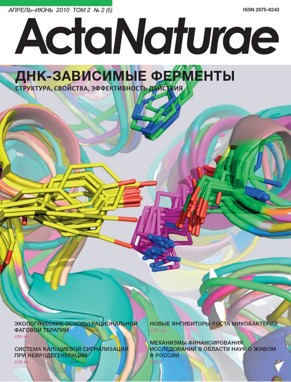
Bioinformatic Analysis, Molecular Modeling of Role of Lys65 Residue in Catalytic Triad of D-aminopeptidase from Ochrobactrum anthropi

Обложка

Цитировать

Полный текст

Аннотация

ABSTRACT A bioinformatic and phylogenetic study has been performed on a family of penicillin-binding proteins including D-aminopeptidases, D-amino acid amidases, DD-carboxypeptidases, and β-lactamases. Significant homology between D-aminopeptidase from Ochrobactrum anthropi and other members of the family has been shown and a number of conserved residues identified as S62, K65, Y153, N155, H287, and G289. Three of those (Ser62, Lys65, and Tyr153) form a catalytic triangle - the proton relay system that activates the generalized nucleophile in the course of catalysis. Molecular modeling has indicated the conserved residue Lys65 to have an unusually low pKa value, which has been confirmed experimentally by a study of the pH-profile of D-aminopeptidase catalytic activity. The resulting data have been used to elucidate the role of Lys65 in the catalytic mechanism of D-aminopeptidase as a general base for proton transfer from catalytic Ser62 to Tyr153, and vice versa, during the formation and hydrolysis of the acylenzyme intermediate.

Полный текст

Bioinformatic Analysis, Molecular Modeling of Role of Lys65 Residue in Catalytic Triad of D-aminopeptidase from Ochrobactrum anthropi
×

Список литературы

  1. Asano Y., Nakazawa A., Kato Y., Kondo K. // J. Biol. Chem. 1989. V. 264. P. 14233-14239.
  2. Bompard-Gilles C., Remaut H., Villeret V., et al. // Structure. 2000. V. 8. P. 971-980.
  3. Asano Y., Kato Y., Yamada A., Kondo K. // Biochemistry. 1992. V. 31. P. 2316-2328.
  4. Okazaki S., Suzuki A., Komeda H., et al. // J. Mol. Biol. 2007. V. 368. P. 79-91.
  5. Massova I., Kollman P. // J. Comput. Chem. 2002. V. 23. P. 1559-1576.
  6. Ke Y., Lin T. // Biophys. Chem. 2005. V. 114. P. 103-113.
  7. Gherman B., Goldberg S., Cornish V., Friesner R. // J. Am. Chem. Soc. 2004. V. 126. P. 7652-7664.
  8. SciDAVis: a free application for scientific data analysis and visualization. http://scidavis.sourceforge.net/
  9. Altschul S., Madden T., Schaffer A., et al. // Nucleic Acids Res. 1997. V. 25. P. 3389-3402.
  10. Notredame C., Higgins D., Heringa J. // J. Mol. Biol. 2000. V. 302. P. 205-217.
  11. Katoh K., Asimenos G., Toh H. // Methods Mol. Biol. 2009. V. 537. P. 39-64.
  12. Do C., Mahabhashyam M., Brudno M., Batzoglou S. // Genome Res. 2005. V. 15. P. 330-340.
  13. Krissinel E., Henrick K. // Acta Cryst. 2004. V. D60. P. 2256-2268.
  14. Konagurthu A., Whisstock J., Stuckey P., Lesk A. // Proteins. 2006. V. 64. P. 559-574.
  15. Felsenstein J. 2005. PHYLIP (Phylogeny Inference Package) version 3.6. Distributed by author. Department of Genome Sciences, University of Washington, Seattle, USA.
  16. Waterhouse A., Procter J., Martin D., Clamp M., Barton G. // Bioinformatics. 2009. V. 25. P. 1189-1191.
  17. The PyMOL Molecular Graphics System, Version 1.0r1, Schrodinger, LLC. http://www.pymol.org/
  18. Crooks G., Hon G., Chandonia J., Brenner S. // Genome Research. 2004. V. 14. P. 1188-1190.
  19. Koonin E., Galperin M. Sequence - Evolution - Function: Computational Approaches in Comparative Genomics. Kluwer Academic Publishers, 2003. P. 461.
  20. Pearl F., Bennett C., Bray J., et al. // Nucleic Acids Res. 2003. V. 31. P. 452-455.
  21. Li H., Robertson A., Jensen J. // Proteins. 2005. V. 61. P. 704-721.

Дополнительные файлы

Доп. файлы
Действие
1. JATS XML

© Khaliullin I.G., Suplatov D.A., Shalaeva D.N., Otsuka M., Asano Y., Svedas V.K., 2010

Creative Commons License
Эта статья доступна по лицензии Creative Commons Attribution 4.0 International License.

Данный сайт использует cookie-файлы

Продолжая использовать наш сайт, вы даете согласие на обработку файлов cookie, которые обеспечивают правильную работу сайта.

О куки-файлах