


Технология гидрогелевых биочипов ИМБ РАН: 30 лет спустя
- Авторы: Грядунов Д.A.1, Шаскольский Б.Л.1, Наседкина T.В.1, Рубина A.Ю.1, Заседателев A.С.1
-
Учреждения:
- Институт молекулярной биологии им. В.А. Энгельгардта РАН
- Выпуск: Том 10, № 4 (2018)
- Страницы: 4-18
- Раздел: Обзоры
- Дата подачи: 17.01.2020
- Дата публикации: 15.12.2018
- URL: https://actanaturae.ru/2075-8251/article/view/10303
- DOI: https://doi.org/10.32607/20758251-2018-10-4-4-18
- ID: 10303
Цитировать

Аннотация
Биологические микрочипы являются аналитическим инструментом, позволяющим реализовать в доступной форме сложные интегративные подходы геномики и протеомики, решать задачи персонализированной медицины - проводить обследование пациента для выявления заболевания задолго до проявлений клинических симптомов, прогнозировать тяжесть протекания патологических и инфекционных процессов, выбирать эффективные и рациональные режимы терапии. Эффективность биочипов обусловлена возможностью параллельного проведения множества специфических реакций и исследования взаимодействий молекул биополимеров, таких, как ДНК, белки, гликаны. Одним из пионеров технологии биологических микрочипов стал Институт молекулярной биологии им. В.А. Энгельгардта Российской академии наук (ИМБ РАН), предложив иммобилизацию молекулярных зондов в трехмерной структуре гидрофильного геля. С момента первых экспериментов по секвенированию с помощью гибридизации на олигонуклеотидных микроматрицах, проведенных 30 лет назад, гидрогелевые биочипы ИМБ РАН прошли долгий и успешный путь от фундаментальных исследований до клинической лабораторной диагностики. Настоящий обзор посвящен обсуждению ключевых аспектов технологии гидрогелевых биочипов и ряду актуальных приложений к мультиплексному анализу ДНК и белковых биомаркеров социально значимых заболеваний, включая молекулярно-генетические, иммунологические и эпидемиологические аспекты патогенеза.
Полный текст
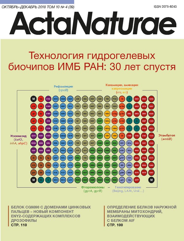
ВВЕДЕНИЕ В последнее десятилетие накоплено большое количество знаний о молекулярных механизмах биохимических процессов, лежащих в основе функционирования живых систем и позволяющих оценивать вероятность возникновения заболевания до проявления у пациента клинических симптомов, прогнозировать тяжесть протекания патологических или инфекционных процессов, выбирать эффективные и рациональные режимы терапии. Решение задач персонифицированной медицины должно включать как масштабный анализ нуклеотидных последовательностей геномов, так и мультиплексные методы количественного определения маркеров патологических состояний. Для одновременного количественного определения последовательностей нуклеиновых кислот (НК) разработано немало методов и технологий, одной из которых, успешно зарекомендовавшей себя в транскрипционном профилировании, сравнительной геномной гибридизации, при одновременной идентификации множества мишеней в геномах человека, растений, микроорганизмов и вирусов, является технология биологических микрочипов (биочипов) [1]. Ключевой элемент биочипа - матрица ячеек, каждая из которых содержит зонд, последовательность которого специфична к фрагменту анализируемого генома. Одновременно проводимые в каждом из элементов биочипа реакции гибридизации и/или амплификации НК обеспечивают параллельную идентификацию разных геномных мишеней, тем самым реализуя принцип многопараметрического анализа биологического образца, что позволяет использовать ДНК-чипы в качестве эффективного молекулярного инструмента выявления клинически значимых маркеров возбудителей и причин социально значимых заболеваний. Биочипы могут также содержать ячейки с иммобилизованными белками или олигосахаридами, причем в каждой отдельной ячейке может быть иммобилизован как индивидуальный зонд, так и их комбинации в зависимости от целей эксперимента. Взаимодействие между различными классами молекул может происходить по типу «рецептор-лиганд», «антиген-антитело», «фермент-субстрат» и т.д. Иммобилизованный лиганд при инкубации с образцом, содержащим анализируемые молекулы, образует специфический комплекс. На этой стадии происходит разделение анализируемых соединений из смеси по их способности к специфическому связыванию с иммобилизованными лигандами, что позволяет анализировать одновременно несколько биологических объектов на одном биочипе, реализуя принцип мультиплексного иммуноанализа. Такой тест необходим для исследований в области протеомики, для диагностики заболеваний, характеризующихся изменением большого числа показателей в сыворотке крови пациента. КЛЮЧЕВЫЕ АСПЕКТЫ АНАЛИЗА НА БИОЧИПАХ В основе проведения анализа на ДНК-микрочипах лежит гибридизация НК. Преимуществами гибридизации являются простота, мультиплексность и воспроизводимость. В отличие от ферментативных реакций, гибридизацию можно проводить в широком диапазоне температур и композиций буферов. В то же время гибридизация не обеспечивает непосредственной амплификации НК и должна сочетаться с технологиями усиления сигналов либо с высокочувствительными инструментами для регистрации дуплексов НК. Поэтому микрочипы используют для прямой количественной детекции РНК, выделенной из больших объемов образца, или для регистрации гибридизационных комплексов, образованных иммобилизованными зондами и фрагментами НК, полученными на предварительной стадии амплификации. Таким образом, чувствительность анализа на ДНК-чипах определяется исходным количеством НК, эффективностью амплификации и способом регистрации комплексов. В случае наиболее распространенной флуоресцентной детекции взаимодействий в элементах биочипа чувствительность метода зависит от анализатора флуоресценции. Теоретически ДНК-чипы должны обеспечивать количественное определение НК [2]. Реальные эксперименты показывают значительные количественные отклонения при оценке экспрессии генов разными платформами микрочипов и даже разными микрочипами одного и того же производителя [3]. Во-первых, кинетика гибридизации нелинейно определяется плотностью зондов на поверхности матрицы, поскольку олигонуклеотиды, иммобилизованные или синтезированные на подложках чипов высокой плотности, неспецифично гибридизуются друг с другом в зависимости от степени гомологии. Во-вторых, на кинетику гибридизации влияют длина и нуклеотидная последовательность целевых молекул ДНК. В-третьих, квантовый выход используемого для регистрации флуорофора зависит как от последовательности прилегающей НК, так и от близости других флуорофоров. В связи с этим экспрессионные микрочипы чаще используются для воспроизводимого анализа относительного содержания НК, чем для точного определения концентрации [4]. Один из ключевых параметров, характеризующих микрочипы - тип субстрата матрицы, - подложки c гидрогелевым покрытием, например, из полиакриламида или агарозы, а также матрицы с функциональными группами, такими, как альдегиды, эпокси- или аминогруппы и др. [5]. В силу гидрофильных свойств полимерные гидрогели являются приоритетным субстратом для иммобилизации биомолекул. Традиционным подходом к изготовлению микрочипов считается покрытие подложки однородным слоем гидрогеля с последующим нанесением зондов либо синтезом олигонуклеотидов in situ. В качестве сшивающих агентов для формирования гидрогелевых подложек используют как синтетические поли-2-гидроксиэтилметакрилат, полиакриламиды, так и несинтетический полимер коллаген [6]. В результате емкость иммобилизации зондов на биочипах возрастает на несколько порядков [7], что позволяет регистрировать сигналы в элементах чипа, в 10-100 раз превышающие сигналы при иммобилизации на планарных матрицах. Принципиальное отличие технологии биочипов, созданной в ИМБ РАН под руководством академика Андрея Дарьевича Мирзабекова (1937-2003), - иммобилизация молекулярных зондов в трехмерных элементах гидрогеля, закрепленных на плоской подложке [8, 9]. Молекулярные зонды - олигонуклеотиды и олигосахариды - модифицируют посредством присоединения амино- или сульфогрупп, используемых далее для ковалентного присоединения в ходе роста полимерной цепи. В то же время зонды белковой природы не нуждаются в специальной модификации, поскольку имеют в своей структуре соответствующие функциональные группы аминокислот. Макропористая структура гидрогелевых элементов формируется за счет сополимеризации мономера - производного метакриловой кислоты, непредельного производного О- или N-замещенного сахара, бифункционального кросс-сшивающего агента и молекулярного зонда. Композиции полимеризационной смеси объемом 0.1 нл наносят на подложку с помощью пинов механического робота (рис. 1). В качестве подложки могут использоваться практически любые носители. Далее под действием ультрафиолетового излучения происходит совместная полимеризация молекулярных зондов с основными компонентами гидрогеля и равномерное встраивание иммобилизуемых соединений в растущую полимерную структуру. Необходимо отметить, что подобраны условия полимеризации молекулярных зондов, позволяющие максимально сохранить их исходную биологическую активность. По завершении полимеризации ячейки гидрогеля, сформировавшиеся на подложке, отмывают и подготавливают к проведению исследований. Эффективность такой иммобилизации составляет 50-80% в зависимости от молекулярного зонда. В зависимости от типа биочипа диаметры гелевых ячеек варьируют от 50 до 300 мкм, а расстояния между ячейками - от 100 до 500 мкм. Количество ячеек на биочипе зависит от задачи и составляет от нескольких десятков до нескольких тысяч. Контроль качества нанесения осуществляется посредством специализированного аппаратно-программного комплекса. Биочипы с отклонением геометрических параметров элементов, не превышающим 10%, а также с отклонениями параметров чипов между сериями, составляющими не более 20%, используют в дальнейших экспериментах [10]. Данные характеристики соответствуют критериям, применяемым для лучших коммерческих микроматриц, производимых компаниями ArrayIt (США) и Schott AG (Германия). С момента первых экспериментов по секвенированию с помощью гибридизации на олигонуклеотидных микроматрицах 30 лет назад [11] гидрогелевые биочипы прошли долгий путь от фундаментальных исследований к клинической лабораторной диагностике. Исследование кинетики и термодинамики гибридизации фрагментов ДНК показало, что использование коротких (до 25 нуклеотидов) зондов обеспечивает эффективную дискриминацию точечных нуклеотидных замен, а иммобилизация в трехмерных гелевых элементах позволяет значительно увеличить интенсивность положительных сигналов и уменьшить статистический разброс по сравнению с двумерными микрочипами [12, 13] (рис. 2). Экспериментально подобраны условия гибридизации, концентрации иммобилизованных зондов, предложены алгоритмы расчета последовательностей зондов, обеспечивающие высокие значения положительного сигнала и дискриминационных отношений [14]. Достигнут значительный прогресс в аналитической чувствительности анализа за счет создания процедуры мультиплексной ПЦР, обеспечивающей одновременную амплификацию десятков фрагментов геномов [15, 16], а также благодаря синтезу новых красителей и оптимизации флуоресцентного маркирования [17, 18]. Впервые были разработаны способы эффективной иммобилизации белковых и гликановых молекул в гидрогеле, предложены методы мультиплексного количественного анализа различных белков в сыворотке крови [19]. Создано несколько поколений универсальных флуоресцентных анализаторов, последнее из которых обеспечивает измерение интенсивности сигналов элементов биочипа при длинах волн от 380 до 850 нм, позволяя проводить качественный и количественный анализ с точностью ± 5% [20]. Созданная в ИМБ РАН универсальная платформа гидрогелевых биочипов позволила разработать, валидировать и внедрить в практику ряд актуальных приложений для многопараметрического анализа биомаркеров социально значимых заболеваний, которые будут детально рассмотрены ниже. АНАЛИЗ СПЕЦИФИЧЕСКИХ ПОСЛЕДОВАТЕЛЬНОСТЕЙ БАКТЕРИАЛЬНЫХ И ВИРУСНЫХ ГЕНОМОВ Необходимость исследования геномов бактерий и вирусов определяется прежде всего высокой социальной значимостью патогенов, к которым зачастую относятся лекарственно-устойчивые возбудители туберкулеза (MTB) и микобактериозов (NTM), вирус гепатита С (ВГС), микроорганизмы группы инфекционно-воспалительных заболеваний репродуктивных органов. Применительно к данным объектам технология гидрогелевых ДНК-биочипов зарекомендовала себя как эффективный инструмент определения профиля детерминант антибиотикорезистентности, внутри- и межвидового генотипирования микроорганизмов и вирусов, необходимого для выбора рациональной терапии и осуществления эпидемиологического надзора. Биочипы в лабораторной диагностике туберкулеза Первой в мире методикой на основе биочипов, разработанной и разрешенной к применению в лабораторной диагностике in vitro, стал набор реагентов «ТБ-Биочип-1» для идентификации 48 мутаций в геноме Mycobacterium tuberculosis, ответственных за устойчивость к рифампицину (RMP) и изониазиду (INH) [21]. Диагностические характеристики метода были оценены на основании результатов десятилетнего (2005-2015 гг.) применения набора «ТБ-Биочип-1» в учреждениях противотуберкулезной службы РФ, Кыргызстана, Азербайджана. Метаанализ 16 публикаций, содержащих данные исследования более 5000 клинических образцов и изолятов с использованием набора «ТБ-Биочип-1» и микробиологических тестов, показал, что диагностическая чувствительность метода при идентификации устойчивого к RMP фенотипа MTB находится в диапазоне 88.8-96.9%, специфичность - 90.3-99.4%; при анализе INH-устойчивых штаммов чувствительность составляет 85.7-96.9%, специфичность - 85.3-98.2%. Совпадение результатов, полученных c помощью набора «ТБ-Биочип-1» и рекомендованных ВОЗ молекулярных тестов Xpert MTB/RIF (Сepheid, США) и Genotype MTBRDplus (Hain Lifescence, Германия), составило 80-90% [22, 23]. Важным результатом применения набора «ТББиочип-1» стала подтвержденная главным внештатным фтизиатром Минздрава РФ оценка клинической эффективности этого набора при лечении пациентов с деструктивным легочным туберкулезом в зависимости от времени коррекции схемы терапии [24]. .При ранней диагностике МЛУ-форм MTB с помощью биочипов число излеченных больных увеличилось по меньшей мере в 3 раза, в отличие от диагностики традиционными методами культивирования [14, 25]. В настоящее время набор «ТБ-Биочип-1» продолжает активно применяться в лабораторной диагностике туберкулеза, оперативно выявляя МЛУ-изоляты и позволяя переводить таких пациентов на другие режимы терапии. Вместе с тем, последовательное накопление мутаций, ассоциированных с резистентностью, привело не только к росту впервые выявленных больных с МЛУ-формами туберкулеза (с ~15% в 2005 г. [21] до ~50% в 2015 г. [26]), но и к появлению изолятов с широкой (ШЛУ) и тотальной устойчивостью ко всем противотуберкулезным препаратам. Для решения данных задач нами разработан метод выявления ДНК MTB с одновременным установлением генотипа эндемичных для РФ штаммов и определением генетических детерминант МЛУ и ШЛУ. Процедура анализа включала мультиплексную ПЦР с адаптерными праймерами и циклической элонгацией с целью одновременной амплификации и флуоресцентного маркирования 17 локусов генома M. tuberculosis с последующей гибридизацией. Биочип, являющийся ключевым компонентом разработанного подхода, позволяет идентифицировать ДНК MTB, определять эндемичные для РФ семейства возбудителя и выявлять суммарно 116 генетических детерминант лекарственной устойчивости к рифампицину, изониазиду, фторхинолонам, инъекционным препаратам (амикацину, канамицину, капреомицину), этамбутолу (EMB) (рис. 3). Клинические испытания метода, проведенные в Санкт-Петербургском НИИ фтизиопульмонологии Минздрава РФ, показали, что значения диагностической чувствительности и специфичности созданной методики по всем препаратам составили 90% и более, за исключением этамбутола [15], чувствительность по которому, равная 89.9%, существенно превосходила опубликованное ранее значение (58%) [27]. Исследование семейств MTB выявило преобладание штаммов генотипа Beijing (73.1%); в меньшей степени были представлены семейства LAM (12.1%), Ural (~7%) и штаммы, относящиеся к ЕвроАмериканской линии (7.2%) (рис. 4). Из всех изолятов, принадлежащих к генотипу Beijing, более 30% составил кластер B0/W148. Установление принадлежности изолята к данному генотипу в 100% означало выявление МЛУ- или ШЛУ-фенотипа, подтверждая клиническую значимость обнаружения данного семейства. Наоборот, изоляты Евро-Американской линии, не входящие в семейства LAM и Ural, были преимущественно ассоциированы с чувствительным фенотипом [15]. Совместно с Московским научно-практическим центром борьбы с туберкулезом Департамента здравоохранения г. Москвы были установлены корреляции между генетическими детерминантами резистентности и значениями минимальных ингибирующих концентраций, характеризующими уровень устойчивости к противотуберкулезному препарату [28]. Крайне важной представляется возможность дифференцированного назначения различных доз противотуберкулезных препаратов, входящих в чрезвычайно ограниченный спектр средств специфической терапии, по результатам анализа детерминант, ассоциированных с разным уровнем устойчивости. На основе созданного метода разработан, испытан, зарегистрирован в Росздравнадзоре и внедрен в клиническую практику набор реагентов «ТБ-ТЕСТ», в настоящее время приходящий на смену наборам серии «ТБ-Биочип». Спектр анализируемых набором «ТБ-ТЕСТ» геномных мишеней по препаратам первого и второго ряда охватывает, как минимум, режимы I-IV химиотерапии больных туберкулезом в соответствии с Приказом Минздрава РФ № 951 от 29.12.2014 г. При этом скорость и возможность анализа респираторного материала позволяет применять набор «ТБ-ТЕСТ» для быстрого скрининга образцов от пациентов с последующей коррекцией режимов терапии и переводом на новейшие противотуберкулезные препараты [29]. С целью рутинного внутривидового генотипирования штаммов микобактерий туберкулезного комплекса разработан набор «СПОЛИГО-БИОЧИП», позволяющий составить генетический профиль каждого изолята MTB, относя его к определенному генотипу [30]. Разработанный подход используется также для дифференциации MTB от вакцинного штамма M. bovis BCG в содержимом холодных абсцессов у детей с поствакцинальными осложнениями. Видоспецифичный полиморфизм гена gyrB микроорганизмов, принадлежащих роду Mycobacterium, позволил сконструировать биочип, обеспечивающий идентификацию 35 различных видов микобактерий [31]. Исследование популяции микобактерий Центрального и Северо-Западного регионов РФ выявило преобладающие в Европейской части РФ виды NTM, такие, как комплекс M. avium (39%), M. fortuitum (17%) и M. xenopi (13%), проявляющие себя при иммуносупрессии, хронической обструктивной болезни легких и ВИЧ-инфекции [31]. Таким образом, совокупность тест-систем для анализа возбудителей туберкулеза и микобактериозов позволяет проводить комплексное исследование материала от больных в рамках универсальной диагностической платформы биочипов в условиях клинической лаборатории. Единый формат анализа, отвечающий современным требованиям, автоматизированная обработка результатов и их интерпретация в виде выдачи конкретных рекомендаций являются основой совершенствования схем терапии туберкулеза, вызываемого лекарственно-устойчивыми формами. Анализ генетических детерминант резистентности возбудителей инфекций репродуктивного тракта (ИРТ) к антимикробным препаратам (АМП) Разнообразие ИРТ, их частое развитие по типу смешанных и лекарственно-устойчивых форм, включающих как облигатные патогены, передающиеся половым путем, так и ряд возбудителей оппортунистических инфекций, существенно затрудняет диагностику данных заболеваний и осложняет выбор стратегии персонализированной терапии. Среди возбудителей ИРТ особое место занимает Neisseria gonorrhoeae. Как и возбудитель туберкулеза, гонококки характеризуются экстраординарной способностью развивать лекарственную устойчивость. В отличие от MTB, N. gonorrhoeae приобретает новые резистентные свойства не только посредством хромосомных мутаций, но и активно использует разнообразные мобильные элементы, а также горизонтальный перенос генов от других видов. Особенно эффективно возбудитель гонореи использует мута16S рРНК (идентификация) gyrA mtrR penA ponA 16S рРНК (детерминанты устойчивости) А ntr4tv ntr6tv blaTEM porB rpsJ parC 23S рРНК Б Рис. 5. Биочип для анализа резистентности возбудителей ИРТ, содержащий зонды, соответствующие видоспецифичному полиморфизму гена 16S рРНК для идентификации возбудителей ИРТ, а также зонды, специфичные к последовательностям генов rrs, rrl, gyrA, parC, mefA, mtrR, nimB-G, penA, ponA, porB, rpsJ, ntr4tv, ntr6tv, blaSHV, blaTEM, tetM, являющихся детерминантами резистентности к АМП. Элементы, содержащие олигонуклеотиды дикого типа, выделены толстым контуром (А). Гибридизационная картина анализа образца ДНК изолята N. gonorrhoeae, содержащего мутации: S91F+D95G в гене gyrA (группа 2), -35delA в промоторе гена mtrR (группа 3), insD345 в гене penA (группа 4), S87R в гене parC (группа 10) (Б) ции, влияющие на проницаемость мембран и увеличивающие активность эффлюксных помп, поскольку эти системы обладают свойством формировать резистентность одновременно ко многим АМП [32]. Разработан биочип (рис. 5А) и метод на его основе для идентификации ДНК 12 различных облигатных и условно-патогенных микроорганизмов с одновременным дифференцированным анализом 39 генетических детерминант устойчивости к β-лактамным антибиотикам, макролидам, аминогликозидам, тетрациклину, спектиномицину, фторхинолонам и нитроимидазолу [33]. Анализ более 500 клинических образцов и изолятов, выделенных в Государственном научном центре дерматовенерологии и косметологии Минздрава России, продемонстрировал высокую чувствительность и специфичность созданного метода при обнаружении ДНК возбудителей ИРТ, а также предсказательную эффективность идентификации маркеров их резистентности к АМП. Исследование тетрациклин-устойчивых штаммов гонококка, выделенных в 2015-2017 гг. в России, показало, что долговременный (с 2003 г.) отказ от использования тетрациклина для лечения гонореи ривел к снижению доли резистентных штаммов в РФ с 75 до 42.6%, что, однако, не позволяет рекомендовать тетрациклины к возвращению в спектр актуальных препаратов для терапии гонококковой инфекции. Впервые в России был охарактеризован тип плазмидного гена tetM, продукт которого приводил к высокому уровню устойчивости к тетрациклину, независимо от присутствия детерминант резистентности хромосомной локализации [34]. Штаммы N. gonorrhoeae содержали множественные мутации в генах penA, ponA, rpsJ, gyrA, parC, mtrR и др. (рис. 5Б), прогностическое значение которых в отношении фенотипической устойчивости существенно возрастало при обнаружении сочетаний генетических детерминант резистентности (рис. 6) [35, 36]. Это обстоятельство вступает в определенное противоречие с тем фактом, что современная терапия гонококковой инфекции основана на преимущественном использовании цефалоспоринов третьего поколения и формально не ведет к «селекционному давлению» по генетическим детерминантам, контролирующим устойчивость к ранее использованным препаратам (пенициллинам, фторхинолонам). Тем самым можно было ожидать элиминации таких мутаций из генома современной популяции N. gonorrhoeae. Возможным объяснением присутствия мутаций представляется многофакторный характер феномена антибиотикорезистентности, в котором ряд ранее мутировавших генов служит основой для следующего витка молекулярной эволюции N. gonorrhoeae. В частности, это относится к генам penA и ponA, мутации в которых оказываются значимыми и в развивающейся устойчивости к цефалоспоринам. Тем самым мы вправе ожидать возникновения устойчивости к современным АМП, в первую очередь, среди мультирезистентных штаммов N. gonorrhoeae, как это имеет место в странах ЕС [37]. Указанное обстоятельство свидетельствует в пользу актуальности непрерывного мониторинга антибиотикорезистентности возбудителя гонореи, одним из инструментов которого сегодня становится технология гидрогелевых биочипов. Определение генотипа и подтипа вируса гепатита С (ВГС) на основе анализа области NS5B вирусного генома Согласно современной классификации, ВГС предложено подразделять на семь основных генотипов и 67 подтипов [38]. Генотип и подтип ВГС являются ключевыми детерминантами при выборе режимов терапии препаратами прямого противовирусного действия (ПППД), действующими на основные мишени жизненного цикла вируса [39]. Точная идентификация генотипа и подтипа ВГС определяет показания к терапии посредством выбора ПППД, продолжительности курса и необходимости назначения рибавирина. Совместно с лабораторией вирусологии госпиталя Университета г. Тулузы (Франция) предложен метод идентификации шести генотипов и 36 подтипов ВГС посредством анализа генотип- и подтипспецифичных последовательностей области NS5B генома ВГС на гидрогелевом биочипе (pис. 7А). Процедура анализа включала амплификацию и флуоресцентное маркирование области NS5B с последующей гибридизацией на биочипе, регистрацией сигналов и их интерпретацией. Пример анализа образца РНК ВГС подтипа 1b с иллюстрацией интерпретации результата приведен на pис. 7Б. С использованием разработанного метода исследованы 345 образцов ВГС в сравнении с «золотым стандартом» генотипирования - секвенированием области NS5B с последующим филогенетическим анализом. Обоими методами был успешно определен генотип всех образцов РНК при 100% корреляции. Совпадение результатов идентификации подтипов получено для 329 из 330 образцов [40]. По своим характеристикам разработанный набор «HCV-Биочип» не уступает технологиям секвенирования, являясь эффективным инструментом рутинного генотипирования, он актуален для оценки ответа на терапию ПППД в зависимости от подтипа ВГС [41, 42]. БИОЧИПЫ ДЛЯ ПЕРСОНАЛИЗИРОВАННОЙ ТЕРАПИИ ОНКОЛОГИЧЕСКИХ БОЛЬНЫХ Молекулярно-генетический анализ химерных генов при лейкозах Выявление структурных перестроек генома с образованием химерных генов в опухолевых клетках костного мозга, особенно у детей, используется в большинстве современных протоколов для разделения пациентов на группы риска и выбора терапии. Тест-система «ЛК-БИОЧИП» разработана и зарегистрирована в Росздравнадзоре для анализа 13 клинически наиболее значимых хромосомных транслокаций при лейкозах [43]. «ЛК-БИОЧИП» применяли для диагностики хромосомных транслокаций у детей в мультицентровых исследованиях по лечению острого лимфобластного лейкоза («ОЛЛ MB-2002» и «ОЛЛ MB-2008») в РФ c 2005 по 2015 г. [44]. В следующей генерации биочипов для диагностики лейкозов представлен расширенный спектр хромосомных транслокаций, включающий еще 9 клинически значимых перестроек t(1;11) MLL/MLLT11, t(1;11) MLL/EPS15, Del1 SIL/TAL1, t(2;5) NPM1/ALK, t(16;21) FUS/ERG, t(1;22) RBM15-MKL1, t(10;11) CALM/AF10, t(17;19) E2A/HLF, t(6;9) DEK/CAN (рис. 8). Тест-система позволяет выявлять в клиническом образце одну опухолевую клетку среди 1000 нормальных со специфичностью не менее 95% [45]. Биочипы для анализа соматических мутаций Выявление соматических мутаций в опухолевой ткани позволяет отобрать для терапии препараты, направленно действующие на данные молекулярные мишени. Доля мутантной ДНК в анализируемом материале зачастую незначительна в силу гетерогенности опухоли или загрязнения образца нормальной тканью. Материалом для молекулярно-генетического исследования обычно служит опухолевая ткань, фиксированная в парафиновых блоках. В результате такого способа хранения опухолевая ДНК подвергается частичной деградации и фрагментации, что накладывает определенные ограничения на использование молекулярно-генетических методов. Емельяновой и соавт. разработан метод анализа соматических мутаций на биочипе с пределом чувствительности выявления мутантной ДНК до 0.5% [46]. С использованием этого подхода были проанализированы соматические мутации при меланоме кожи, прорыв в лечении которой в последние годы связан с применением таргетных препаратов, действующих на специфические молекулярные мишени, и иммунотерапии, эффективность которой во многом зависит от генотипа опухоли. Основой этого метода является биочип для определения 39 клинически значимых соматических мутаций в генах BRAF, NRAS, KIT, GNAQ, GNA11, MAP2K1 и MAP2K2 (рис. 9). Разработанным методом было протестировано 253 клинических образца меланомы. Выявлены разнообразные мутации в генах BRAF (51.0%), NRAS (17.8%), KIT (2.4%), GNAQ (1.6%), GNA11 (0.8%), MAP2K1 (0.8%). Подход позволяет эффективно выявлять клинически значимые соматические мутации и делать выбор в пользу определенного таргетного препарата у 70% больных меланомой [47]. МУЛЬТИПЛЕКСНЫЙ ИММУНОАНАЛИЗ НА БИОЧИПАХ В зависимости от клинической задачи выделяют два основных типа мультиплексного иммуноанализа: определение различных индивидуальных антигенов в образце или выявление антител, циркулирующих в сыворотке крови. В первом случае биочип содержит панель иммобилизованных антител, каждое из которых специфично взаимодействует с конкретным определяемым антигеном. Во втором, в структуру биочипа включены иммобилизованные лиганды белковой или иной природы, специфично связывающиеся с иммуноглобулинами в составе образца. Примером разработанного в ИМБ РАН метода первого типа может служить разработанная совместно с сотрудниками ИБХ РАН под руководством академика Е.В. Гришина тест-система для количественного определения ряда биотоксинов [48]. Мультиплексный анализ онкомаркеров на биочипах Поиску клинически значимых биомаркеров, обладающих высокой чувствительностью и специфичностью в отношении опухолей конкретного вида, посвящено огромное число исследований. Повысить диагностическую эффективность позволяет одновременное определение нескольких онкомаркеров. Вместе с тем, такой подход не обеспечивает своевременного выявления некоторых опухолей. Так, для диагностики колоректального рака (КРР) in vitro рекомендовано исследование сывороточных онкомаркеров РЭА и СА 19-9. В то же время результаты масштабных клинических исследований свидетельствуют, что данные биомаркеры выявляют заболевание в основном на поздних стадиях (III и IV) и обладают клинической значимостью только для мониторинга терапии [49]. Большинство КРРассоциированных маркеров являются гликопротеинами или карбогидратными антигенами, они содержат О- либо N-гликосайты [50]. Изменения в гликозилировании данных маркеров приводят к изменению уровней соответствующих антител, что можно детектировать с помощью мультиплексного анализа на биочипах. Совместно с сотрудниками лаборатории углеводов ИБХ РАН разработан подход, основанный на одновременном анализе белковых серологических онкомаркеров и антител различных классов к онкоассоциированным гликанам. Сконструирован комбинированный биочип, элементы которого содержали гликаны, а также антитела к онкомаркерам КРР. Содержание антител к гликанам в сыворотке крови определяли с помощью флуоресцентного анализа на биочипах (рис. 10). В результате исследований, проведенных совместно с МНИОИ им. П.А. Герцена, выявлены комбинации (сигнатуры), состоящие из уровней антител к некоторым опухолеассоциированным гликанам и значений концентраций основных онкомаркеров, позволяющие достоверно отличить больных КРР и здоровых доноров [51]. Показано, что одновременное использование белковой и гликановой сигнатур позволяет выявлять КРР у пациентов надежнее, чем традиционная пара онкомаркеров РЭA + CA 19-9. Чувствительность метода составила 88%, специфичность - 98%, в то время как комбинация РЭА и СА 19-9 позволила выявить КРР в 80% случаев с чувствительностью 21%. Анализ аллергенспецифических иммуноглобулинов (Ig) на биочипе Основными маркерами аллергии на сегодняшний день являются специфические иммуноглобулины класса Е, которые определяют протекание аллергической реакции по механизму первого типа (анафилаксия, отек Квинке). Иммуноглобулины других классов также могут принимать участие в аллергических реакциях. Так, специфические иммуноглобулины класса IgG4 (sIgG4) играют определенную роль в развитии толерантности - отсутствии клинических проявлений на определенные аллергены при наличии сенсибилизации к этим аллергенам. Несмотря на то что sIgG4 не относится к диагностическим маркерам, определение этого параметра важно для оценки соотношения sIgE/sIgG4, которое показывает эффективность проведения специфической иммунотерапии. sIgG4 играют роль блокирующих антител, которые препятствуют развитию аллергических реакций по первому типу [52]. Разработанная в ИМБ РАН совместно с компанией DR. FOOKE Laboratorien GmbH (Германия) тестсистема «Аллергобиочип» (рис. 11) предназначена для проведения параллельного анализа панелей sIgE и sIgG4 к аллергенам следующих классов: пыльцевые (деревья и кустарники), пыльцевые (сорные травы и цветы), пыльцевые (травы и злаки), эпидермальные, яды насекомых, клещевые, пищевые, грибковые. Тест-система представляет собой адаптацию твердофазного иммунохимического анализа с флуоресцентной регистрацией сигналов в формате биочипа. Тестирование метода с использованием более 2000 образцов сывороток крови пациентов с аллергией и здоровых доноров показало эффективность аллергобиочипа для выявления гиперчувствительности типа I. Определены пороги детекции для sIgE и sIgG4, чувствительность и специфичность анализа, диапазон измерения, составивший 0.35-100 МЕ/мл для sIgE и 100-2500 нг/мл для sIgG4 [53, 54]. Проведено эпидемиологическое исследование модельной популяции детей средней полосы России (800 пациентов ДГКБ им. Филатова в возрасте от 0 до 16 лет и 50 здоровых доноров) с целью оценки частот встречаемости сенсибилизации к аллергенам различных классов в зависимости от возраста [55]. Получены профили уровней аллергенспецифических sIgE и sIgG4 в каждом из образцов. Обнаружено, что среди ингаляционных аллергенов сенсибилизацию наиболее часто вызывают пыльца березы и эпителий кошки, в то время как среди пищевых аллергенов sIgE-ответ чаще всего связан с аллергенами куриного яйца и молока (рис. 12). Продукцию sIgG4 в основном вызывают пищевые аллергены, особенно яичный белок. С возрастом доля пациентов с повышенным уровнем sIgE к ингаляционным аллергенам увеличивается, при снижении доли пациентов, сенсибилизированных к большинству пищевых аллергенов, за исключением аллергенов моркови, яблока и персика. ЗАКЛЮЧЕНИЕ С момента первой публикации по секвенированию посредством гибридизации с иммобилизованными октануклеотидными зондами [56] прошло 30 лет. За этот период сотрудниками лаборатории биологических микрочипов ИМБ РАН выстроена универсальная методология многопараметрического анализа белковых и ДНК-маркеров при статистических и клинических исследованиях больших серий биологических образцов различной природы. Создана и сертифицирована по международному стандарту ISO 13485 производственная линия по изготовлению гидрогелевых биочипов мощностью до 1 млн в год. ИМБ РАН получено 12 регистрационных удостоверений Росздравнадзора на разработанные медицинские изделия - наборы реагентов и оборудование для диагностики in vitro с использованием гидрогелевых биочипов. В свете стремительного развития технологий ПЦР и платформ секвенирования следующего поколения ДНК-микрочипы столкнулись с серьезной конкуренцией. Сегодня они занимают промежуточную нишу, подпираемую снизу разнообразными тестами на основе амплификации НК, и испытывают все большее давление сверху со стороны высокопроизводительных технологий секвенирования. В нашем случае иммобилизация любых типов биомолекул в гидрогеле и возможность проведения в нем ферментативных реакций [57], в том числе и изотермических [58], делает возможным создание портативных биосенсоров нового поколения. Так, гидрогелевые элементы могут стать платформой для совместной иммобилизации геномных редакторов - нуклеаз Cas13 и Cas12a, вместе с направляющими и детектирующими молекулами РНК/ДНК [59]. «Программируемая» работа нуклеаз, дополненная при необходимости изотермической амплификацией, в сочетании с созданными микрофлюидными системами для выделения НК [60], позволит создать высокочувствительные CRISPR-биосенсоры, применение которых будет возможно в полевых условиях. Такие комплексные автономные системы, содержащие платформу гидрогелевых биочипов и согласованные с ней поддерживающие модули, позволят получать результаты быстрее, информативнее и точнее, чем в настоящее время, и будут играть ключевую роль в персонализированной медицине будущего. В настоящее время более 2000 пациентов ДГКБ им. Филатова обследованы с использованием аллергобиочипов. Помимо выраженного экономического эффекта, обусловленного форматом «один образец - один анализ на чипе», предлагаемый подход позволяет использовать всего 100 мкл сыворотки крови для выявления аллергена, вызывающего у ребенка выраженную реакцию. При обследовании детей раннего возраста (несколько месяцев) такое минимальное количество образца представляется существенным преимуществом. Перспективным представляется создание белковых биочипов для дифференциальной диагностики ревматологических заболеваний и других нарушений в работе иммунной системы. Многообещающим выглядит также расширение биочипа для анализа предиктивных маркеров онкозаболеваний. Мы надеемся, что развитие нового подхода на основе комплексного анализа сигнатур позволит решить эту непростую задачу. Таким образом, технология гидрогелевых биочипов уже сегодня является эффективным инструментом персонализированной медицины, выполняя молекулярное профилирование множества клинически значимых маркеров возбудителей и причин социально значимых заболеваний, спасая жизнь сотням больных и обеспечивая экономию бюджетных средств при лечении.
Об авторах
Д. A. Грядунов
Институт молекулярной биологии им. В.А. Энгельгардта РАН
Автор, ответственный за переписку.
Email: grad@biochip.ru
Россия
Б. Л. Шаскольский
Институт молекулярной биологии им. В.А. Энгельгардта РАН
Email: grad@biochip.ru
Россия
T. В. Наседкина
Институт молекулярной биологии им. В.А. Энгельгардта РАН
Email: grad@biochip.ru
Россия
A. Ю. Рубина
Институт молекулярной биологии им. В.А. Энгельгардта РАН
Email: grad@biochip.ru
Россия
A. С. Заседателев
Институт молекулярной биологии им. В.А. Энгельгардта РАН
Email: grad@biochip.ru
Россия
Список литературы
- Marzancola M.G., Sedighi A., Li P.C. // Methods Mol. Biol. 2016, V.1368, P.161-178
- Rosenfeld S. // Gene Regul. Syst. Bio. 2010, V.4, P.61-73
- Eklund A.C., Szallasi Z. // Genome Biol. 2008, V.9, №2, P.26
- Salazar R., Roepman P., Capella G., Moreno V., Simon I., Dreezen C., Lopez-Doriga A., Santos C., Marijnen C., Westerga J. // J. Clin. Oncol. 2011, V.29, №1, P.17-24
- Damin F., Galbiati S., Ferrari M., Chiari M. // Biosens. Bioelectron. 2016, V.78, P.367-733
- Le Goff G.C., Srinivas R.L., Hill W.A., Doyle P.S. // Eur. Polym. J. 2015, V.72, P.386-412
- Beyer A., Pollok S., Berg A., Weber K., Popp J. // Macromol. Biosci. 2014, V.14, №6, P.889-898
- Yershov G., Barsky V., Belgovskiy A., Kirillov E., Kreindlin E., Ivanov I., Parinov S., Guschin D., Drobishev A., Dubiley S. // Proc. Natl. Acad. Sci. USA. 1996, V.93, №10, P.4913-4918
- Rubina A.Y., Pan’kov S.V., Dementieva E.I., Pen’kov D.N., Butygin A.V., Vasiliskov V.A., Chudinov A.V., Mikheikin A.L., Mikhailovich V.M., Mirzabekov A.D. // Anal. Biochem. 2004, V.325, №1, P.92-106
- Rubina A.Y., Kolchinsky A., Makarov A.A., Zasedatelev A.S. // Proteomics. 2008, V.8, №4, P.817-831
- Khrapko K.R., Lysov Yu P., Khorlyn A.A., Shick V.V., Florentiev V.L., Mirzabekov A.D. // FEBS Lett. 1989, V.256, №1-2, P.118-122
- Sorokin N.V., Chechetkin V.R., Livshits M.A., Pan’kov S.V., Donnikov M.Y., Gryadunov D.A., Lapa S.A., Zasedatelev A.S. // J. Biomol. Struct. Dyn. 2005, V.22, №6, P.725-734
- Pan’kov S.V., Chechetkin V.R., Somova O.G., Antonova O.V., Moiseeva O.V., Prokopenko D.V., Yurasov R.A., Gryadunov D.A., Chudinov A.V. // J. Biomol. Struct. Dyn. 2009, V.27, №2, P.235-244
- Gryadunov D., Mikhailovich V., Lapa S., Roudinskii N., Donnikov M., Pan’kov S., Markova O., Kuz’min A., Chernousova L., Skotnikova O. // Clin. Microbiol. Infect. 2005, V.11, №7, P.531-539
- Zimenkov D.V., Kulagina E.V., Antonova O.V., Zhuravlev V.Y., Gryadunov D.A. // J. Antimicrob. Chemother. 2016, V.71, №6, P.1520-1531
- Zimenkov D.V., Antonova O.V., Kuz’min A.V., Isaeva Y.D., Krylova L.Y., Popov S.A., Zasedatelev A.S., Mikhailovich V.M., Gryadunov D.A. // BMC Infect. Dis. 2013, V.13, P.240
- Shershov V.E., Lapa S.A., Kuznetsova V.E., Spitsyn M.A., Guseinov T.O., Polyakov S.A., Stomahin A.A., Zasedatelev A.S., Chudinov A.V. // J. Fluoresc. 2017, V.27, №6, P.2001-2016
- Zasedateleva O.A., Vasiliskov V.A., Surzhikov S.A., Kuznetsova V.E., Shershov V.E., Guseinov T.O., Smirnov I.P., Yurasov R.A., Spitsyn M.A., Chudinov A.V. // Nucleic Acids Research 2018, V.46, №12, P.e73
- Rubina A.Y., Dementieva E.I., Stomakhin A.A., Darii E.L., Pan’kov S.V., Barsky V.E., Ivanov S.M., Konovalova E.V., Mirzabekov A.D. // Biotechniques. 2003, V.34, №5, P.1008-1022
- Lysov Y., Barsky V., Urasov D., Urasov R., Cherepanov A., Mamaev D., Yegorov Y., Chudinov A., Surzhikov S., Rubina A. // Biomed. Opt. Express. 2017, V.8, №11, P.4798-4810
- Gryadunov D., Mikhailovich V., Lapa S., Roudinskii N., Donnikov M., Pan’kov S., Markova O., Kuz’min A., Chernousova L., Skotnikova O. // Clin. Microbiol. Infect. 2005, V.11, №7, P.531-539
- Kurbatova E.V., Kaminski D.A., Erokhin V.V., Volchenkov G.V., Andreevskaya S.N., Chernousova L.N., Demikhova O.V., Ershova J.V., Kaunetis N.V., Kuznetsova T.A. // Eur. J. Clin. Microbiol. Infect. Dis. 2013, V.32, №6, P.735-743
- Nosova E., Krasnova M.A., Galkina K., Makarova M.V., Litvinov V.I., Moroz A.M. // Mol. Biol. (Mosk). 2013, V.47, №2, P.267-274
- Vasil’eva I.A., Samoilova A.G., Ergeshov A.E., Bagdasarian T.R., Chernousova L.N. // Vestn. Ross. Akad. Med. Nauk. 2012, V.67, №11, P.9-14
- Daurov R.B., Vasilyeva I.A., Perfilyev A.V., Chernousova L.N., Kuzmin A.V., Glazkova N.A. // Tuberk. Bolezni Legkih. 2010, V.87, №4, P.10-13
- Mokrousov I., Vyazovaya A., Solovieva N., Sunchalina T., Markelov Y., Chernyaeva E., Melnikova N., Dogonadze M., Starkova D., Vasilieva N. // BMC Microbiol. 2015, V.15, P.279
- Koser C.U., Bryant J.M., Comas I., Feuerriegel S., Niemann S., Gagneux S., Parkhill J., Peacock S.J. // J. Antimicrob. Chemother. 2014, V.69, №8, P.2298-2299
- Nosova E.Y., Zimenkov D.V., Khakhalina A.A., Isakova A.I., Krylova L.Y., Makarova M.V., Galkina K.Y., Krasnova M.A., Safonova S.G., Litvinov V.I. // PLoS One. 2016, V.11, №11, e0167093
- Zimenkov D.V., Nosova E.Y., Kulagina E.V., Antonova O.V., Arslanbaeva L.R., Isakova A.I., Krylova L.Y., Peretokina I.V., Makarova M.V., Safonova S.G. // J. Antimicrob. Chemother. 2017, V.72, №7, P.1901-1906
- Bespyatykh J.A., Zimenkov D.V., Shitikov E.A., Kulagina E.V., Lapa S.A., Gryadunov D.A., Ilina E.N., Govorun V.M. // Infect. Genet. Evol. 2014, V.26, P.41-46
- Zimenkov D.V., Kulagina E.V., Antonova O.V., Krasnova M.A., Chernyaeva E.N., Zhuravlev V.Y., Kuz’min A.V., Popov S.A., Zasedatelev A.S., Gryadunov D.A. // J. Clin. Microbiol. 2015, V.53, №4, P.1103-1114
- Shaskolskiy B., Dementieva E., Leinsoo A., Runina A., Vorobyev D., Plakhova X., Kubanov A., Deryabin D., Gryadunov D. // Front. Microbiol. 2016, V.7, P.747
- Leinsoo A.T., Shaskol’skii B.L., Dement’eva E.I., Gryadunov D.A., Kubanov A.A., Chestkov A.V., Obraztsova O.A., Shpilevaya M.V., Deryabin D.G. // Bull. Exp. Biol. Med. 2017, V.164, №1, P.54-60
- Shaskolskiy B., Dementieva E., Leinsoo A., Petrova N., Chestkov A., Kubanov A., Deryabin D., Gryadunov D. // Infect. Genet. Evol. 2018, V.63, P.236-242
- Kubanov A., Vorobyev D., Chestkov A., Leinsoo A., Shaskolskiy B., Dementieva E., Solomka V., Plakhova X., Gryadunov D., Deryabin D. // BMC Infect. Dis. 2016, V.16, P.389
- Kubanov A.A., Leinsoo A.T., Chestkov A.V., Dementieva E.I., Shaskolskiy B.L., Solomka V.S., Gryadunov D.A., Deryabin D.G. // Mol. Biol. (Mosk). 2017, V.51, №3, P.431-441
- Harris S.R., Cole M.J., Spiteri G., Sanchez-Buso L., Golparian D., Jacobsson S., Goater R., Abudahab K., Yeats C.A., Bercot B. // Lancet. Infect. Dis. 2018, V.18, №7, P.758-768
- Welzel T.M., Bhardwaj N., Hedskog C., Chodavarapu K., Camus G., McNally J., Brainard D., Miller M.D., Mo H., Svarovskaia E. // J. Hepatol. 2017, V.67, №2, P.224-236
- Manns M.P., Buti M., Gane E., Pawlotsky J.M., Razavi H., Terrault N., Younossi Z. // Nat. Rev. Dis. Primers. 2017, V.3, P.17006
- Gryadunov D., Nicot F., Dubois M., Mikhailovich V., Zasedatelev A., Izopet J. // J. Clin. Microbiol. 2010, V.48, №11, P.3910-3917
- Rodriguez-Frias F., Nieto-Aponte L., Gregori J., Garcia-Cehic D., Casillas R., Tabernero D., Homs M., Blasi M., Vila M., Chen Q. // Clin. Microbiol. Infect. 2017, V.23, №10, 10.1016/j.cmi.2017.02.007
- Soria M.E., Gregori J., Chen Q., Garcia-Cehic D., Llorens M., de Avila A.I., Beach N.M., Domingo E., Rodriguez-Frias F., Buti M. // BMC Infect. Dis. 2018, V.18, №1, P.446
- Nasedkina T.V., Guseva N.A., Gra O.A., Mityaeva O.N., Chudinov A.V., Zasedatelev A.S. // Mol. Diagn. Ther. 2009, V.13, №2, P.91-102
- Nasedkina T.V., Ikonnikova A.Y., Tsaur G.A., Karateeva A.V., Ammour Y.I., Avdonina M.A., Karachunskii A.I., Zasedatelev A.S. // Mol. Biol. (Mosk). 2016, V.50, №6, P.968-977
- Ikonnikova A.Yu., Fesenko D.O., Karateeva A.V., Zasedatelev A.S., Nasedkina T.V. // Patent 2639513. Russia. C12N 15/00. 2017, 2639513
- Emelyanova M., Arkhipova K., Mazurenko N., Chudinov A., Demidova I., Zborovskaya I., Lyubchenko L., Zasedatelev A., Nasedkina T. // Appl. Immunohistochem. Mol. Morphol. 2015, V.23, №4, P.255-265
- Emelyanova M., Ghukasyan L., Abramov I., Ryabaya O., Stepanova E., Kudryavtseva A., Sadritdinova A., Dzhumakova C., Belysheva T., Surzhikov S. // Oncotarget. 2017, V.8, №32, P.52304-52320
- Rubina A.Y., Filippova M.A., Feizkhanova G.U., Shepeliakovskaya A.O., Sidina E.I., Boziev Kh.M., Laman A.G., Brovko F.A., Vertiev Y.V., Zasedatelev A.S. // Anal. Chem. 2010, V.82, №21, P.8881-8889
- Duffy M.J., Lamerz R., Haglund C., Nicolini A., Kalousova M., Holubec L., Sturgeon C. // Int. J. Cancer. 2014, V.134, №11, P.2513-2522
- Kufe D.W. // Nat. Rev. Cancer. 2009, V.9, №12, P.874-885
- Butvilovskaya V.I., Popletaeva S.B., Chechetkin V.R., Zubtsova Z.I., Tsybulskaya M.V., Samokhina L.O., Vinnitskii L.I., Ragimov A.A., Pozharitskaya E.I., Grigoreva G.A. // Cancer. Med. 2016, V.5, №7, P.1361-1372
- Vazquez-Ortiz M., Pascal M., Jimenez-Feijoo R., Lozano J., Giner M.T., Alsina L., Martin-Mateos M.A., Plaza A.M. // Clin. Exp. Allergy. 2014, V.44, №4, P.579-588
- Feyzkhanova G.U., Filippova M.A., Talibov V.O., Dementieva E.I., Maslennikov V.V., Reznikov Y.P., Offermann N., Zasedatelev A.S., Rubina A.Y., Fooke-Achterrath M. // J. Immunol. Methods. 2014, V.406, P.51-57
- Feyzkhanova G., Voloshin S., Smoldovskaya O., Arefieva A., Filippova M., Barsky V., Pavlushkina L., Butvilovskaya V., Tikhonov A., Reznikov Y. // Clin. Proteomics. 2017, V.14, P.1
- Voloshin S., Smoldovskaya O., Feyzkhanova G., Arefieva A., Pavlushkina L., Filatova T., Butvilovskaya V., Filippova M., Lysov Y., Shcherbo S. // PLoS One. 2018, V.13, №3, e0194775
- Lysov Iu P., Florent’ev V.L., Khorlin A.A., Khrapko K.R., Shik V.V. // Dokl. Akad. Nauk SSSR. 1988, V.303, №6, P.1508-1511
- Khodakov D.A., Zakharova N.V., Gryadunov D.A., Filatov F.P., Zasedatelev A.S., Mikhailovich V.M. // Biotechniques. 2008, V.44, №2, P.241-248
- Kashkin K.N., Strizhkov B.N., Griadunov D.A., Surzhikov S.A., Grechishnikova I.V., Kreindlin E., Chupeeva V.V., Evseev K.B., Turygin A., Mirzabekov A.D. // Mol. Biol. (Mosk). 2005, V.39, №1, P.30-39
- Gootenberg J.S., Abudayyeh O.O., Kellner M.J., Joung J., Collins J.J., Zhang F. // Science. 2018, V.360, №6387, P.439-444
- Mamaev D., Shaskolskiy B., Dementieva E., Khodakov D., Yurasov D., Yurasov R., Zimenkov D., Mikhailovich V., Zasedatelev A., Gryadunov D. // Biomed. Microdevices. 2015, V.17, P.18
Дополнительные файлы

